Download

1 / 54
540 likes | 773 Vues
This overview explores the fundamental concepts of cell theory, emphasizing that cells are the basic structural and functional units of life. It discusses how organismal functions are dependent on both individual cell activities and their collective operations. The role of specific subcellular structures and the diversity of cell types—such as erythrocytes and nerve cells—are highlighted. Additionally, it details the plasma membrane's composition, including lipids and proteins, and outlines their essential functions such as transport, signaling, and cellular attachment.

E N D
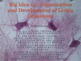
3 Cells: The Living Units:
Cell Theory • Cell - structural and functional unit of life • Organismal functions depend on individual and collective cell functions • Biochemical activities of cells dictated by their shapes or forms, and specific subcellular structures • Continuity of life has cellular basis © 2013 Pearson Education, Inc.
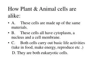
Figure 3.1 Cell diversity. Erythrocytes Fibroblasts Epithelial cells Cells that connect body parts, form linings, or transport gases Skeletal muscle cell Smooth muscle cells Cells that move organs and body parts Macrophage Fat cell Cell that stores nutrients Cell that fights disease Nerve cell Cell that gathers information and controls body functions Sperm Cell of reproduction © 2013 Pearson Education, Inc.
Figure 3.3 The plasma membrane. Extracellular fluid (watery environment outside cell) Polar head of phospholipid molecule Cholesterol Glycolipid Glyco- protein Nonpolar tail of phospholipid molecule Glycocalyx (carbohydrates) Lipid bilayer containing proteins Outward-facing layer of phospholipids Inward-facing layer of phospholipids Cytoplasm (watery environment inside cell) Filament of cytoskeleton Integral proteins Peripheral proteins © 2013 Pearson Education, Inc.
Membrane Lipids • 75% phospholipids (lipid bilayer) • Phosphate heads: polar and hydrophilic • Fatty acid tails: nonpolar and hydrophobic (Review Fig. 2.16b) • 5% glycolipids • Lipids with polar sugar groups on outer membrane surface • 20% cholesterol • Increases membrane stability © 2013 Pearson Education, Inc.
Membrane Proteins • Allow communication with environment • ½ mass of plasma membrane • Most specialized membrane functions • Some float freely • Some tethered to intracellular structures • Two types: • Integral proteins; peripheral proteins © 2013 Pearson Education, Inc.
Membrane Proteins • Integral proteins • Firmly inserted into membrane (most are transmembrane) • Have hydrophobic and hydrophilic regions • Can interact with lipid tails and water • Functions? PLAY Animation: Transport Proteins © 2013 Pearson Education, Inc.
Membrane Proteins • Peripheral proteins • Loosely attached to integral proteins • Include filaments on intracellular surface for membrane support • Functionss? enzymes; motor proteins for shape change during cell division and muscle contraction; cell-to-cell connections PLAY Animation: Structural Proteins PLAY Animation: Receptor Proteins © 2013 Pearson Education, Inc.
Figure 3.3 The plasma membrane. Extracellular fluid (watery environment outside cell) Polar head of phospholipid molecule Cholesterol Glycolipid Glyco- protein Nonpolar tail of phospholipid molecule Glycocalyx (carbohydrates) Lipid bilayer containing proteins Outward-facing layer of phospholipids Inward-facing layer of phospholipids Cytoplasm (watery environment inside cell) Filament of cytoskeleton Integral proteins Peripheral proteins © 2013 Pearson Education, Inc.
Six Functions of Membrane Proteins • Transport • Receptors for signal transduction • Attachment to cytoskeleton and extracellular matrix © 2013 Pearson Education, Inc.
Figure 3.4a Membrane proteins perform many tasks. Transport • A protein (left) that spans the membrane may provide a hydrophilic channel across the membrane that is selective for a particular solute. • Some transport proteins (right) hydrolyze ATP as an energy source to actively pump substances across the membrane. PLAY Animation: Transport Proteins © 2013 Pearson Education, Inc.
Figure 3.4b Membrane proteins perform many tasks. Receptors for signal transduction Signal • A membrane protein exposed to the outside of the cell may have a binding site that fits the shape of a specific chemical messenger, such as a hormone. • When bound, the chemical messenger may cause a change in shape in the protein that initiates a chain of chemical reactions in the cell. Receptor PLAY Animation: Receptor Proteins © 2013 Pearson Education, Inc.
Figure 3.4c Membrane proteins perform many tasks. Attachment to the cytoskeleton and extracellular matrix • Elements of the cytoskeleton (cell's internal supports) and the extracellular matrix (fibers and other substances outside the cell) may anchor to membrane proteins, which helps maintain cell shape and fix the location of certain membrane proteins. • Others play a role in cell movement or bind adjacent cells together. PLAY Animation: Structural Proteins © 2013 Pearson Education, Inc.
Six Functions of Membrane Proteins • Enzymatic activity • Intercellular joining • Cell-cell recognition © 2013 Pearson Education, Inc.
Figure 3.4d Membrane proteins perform many tasks. Enzymatic activity Enzymes • A membrane protein may be an enzyme with its active site exposed to substances in the adjacent solution. • A team of several enzymes in a membrane may catalyze sequential steps of a metabolic pathway as indicated (left to right) here. PLAY Animation: Enzymes © 2013 Pearson Education, Inc. Figure 3.4d
Figure 3.4e Membrane proteins perform many tasks. Intercellular joining • Membrane proteins of adjacent cells may be hooked together in various kinds of intercellular junctions. • Some membrane proteins (cell adhesion molecules or CAMs) of this group provide temporary binding sites that guide cell migration and other cell-to-cell interactions. CAMs © 2013 Pearson Education, Inc.
Figure 3.4f Membrane proteins perform many tasks. Cell-cell recognition • Some glycoproteins (proteins bonded to short chains of sugars) serve as identification tags that are specifically recognized by other cells. Glycoprotein © 2013 Pearson Education, Inc.
Membrane Transport • Concentration = grams of solutes/100 ml water = % or osmoles • Concentration gradient – difference in concentration • Equilibrium – no difference in concentration Mickey Dufilho
Types of Membrane Transport • Passive processes • No cellular energy (ATP) required • Substance moves down its concentration gradient • Active processes • Energy (ATP) required • Occurs only in living cell membranes © 2013 Pearson Education, Inc.
Passive Processes • Two types of passive transport • Diffusion • Simple diffusion • Carrier- and channel-mediated facilitated diffusion • Osmosis • Filtration • Usually across capillary walls © 2013 Pearson Education, Inc.
Passive Processes: Diffusion • Collisions cause molecules to move down or with their concentration gradient • Difference in concentration between two areas • Speed influenced by molecule size and temperature • Molecule will passively diffuse through membrane if • It is lipid soluble, small enough to pass through membrane channels, or assisted by carrier molecule © 2013 Pearson Education, Inc.
Passive Processes: Simple Diffusion • Nonpolar lipid-soluble (hydrophobic) substances diffuse directly through the phospholipid bilayer Mickey Dufilho
Passive Processes: Facilitated Diffusion • Certain lipophobic molecules (e.g., glucose, amino acids, and ions) transported passively by • Binding to protein carriers • Moving through water-filled channels © 2013 Pearson Education, Inc.
Passive Processes: Facilitated Diffusion Mickey Dufilho
Passive Processes: Osmosis • Movement of solvent (e.g., water) across selectively permeable membrane • Water diffuses through plasma membranes • Through lipid bilayer • Through specific water channels called aquaporins (AQPs) • Occurs when water concentration different on the two sides of a membrane © 2013 Pearson Education, Inc.
Figure 3.7d Diffusion through the plasma membrane. Water molecules Lipid bilayer Aquaporin Osmosis, diffusion of a solvent such as water through a specific channel protein (aquaporin) or through the lipid bilayer © 2013 Pearson Education, Inc.
Passive Processes: Osmosis • Water concentration varies with number of solute particles because solute particles displace water molecules • Osmolarity - Measure of total concentration of solute particles • Water moves by osmosis until hydrostatic pressure (back pressure of water on membrane) and osmotic pressure (tendency of water to move into cell by osmosis) equalize © 2013 Pearson Education, Inc.
Figure 3.8a Influence of membrane permeability on diffusion and osmosis. Membrane permeable to both solutes and water Solute and water molecules move down their concentration gradients in opposite directions. Fluid volume remains the same in both compartments. Right compartment: Left compartment: Solution with greater osmolarity Solution with lower osmolarity Both solutions have the same osmolarity: volume unchanged Solute Solute molecules (sugar) Freely permeable membrane © 2013 Pearson Education, Inc.
Figure 3.8b Influence of membrane permeability on diffusion and osmosis. Membrane permeable to water, impermeable to solutes Solute molecules are prevented from moving but water moves by osmosis. Volume increases in the compartment with the higher osmolarity. Both solutions have identical osmolarity, but volume of the solution on the right is greater because only water is free to move Left compartment Right compartment Solute molecules (sugar) Selectively permeable membrane © 2013 Pearson Education, Inc.
Tonicity • Tonicity: Ability of solution to alter cell's water volume • Isotonic: Solution with same non-penetratingsolute concentration as cytosol • Hypertonic: Solution with higher non-penetrating solute concentration than cytosol • Hypotonic: Solution with lower non-penetrating solute concentration than cytosol © 2013 Pearson Education, Inc.
Figure 3.9 The effect of solutions of varying tonicities on living red blood cells. Hypertonic solutions Isotonic solutions Hypotonic solutions Cells retain their normal size and shape in isotonic solutions (same solute/water concentration as inside cells; water moves in and out). Cells lose water by osmosis and shrink in a hypertonic solution (contains a higher concentration of solutes than are present inside the cells). Cells take on water by osmosis until they become bloated and burst (lyse) in a hypotonic solution (contains a lower concentration of solutes than are present inside cells). © 2013 Pearson Education, Inc.
Table 3.1 Passive Membrane Transport Processes © 2013 Pearson Education, Inc.
Passive Membrane Transport: Filtration • The passage of water and solutes through a membrane by hydrostatic pressure • Pressure gradient pushes solute-containing fluid from a higher-pressure area to a lower-pressure area • Does not occur into or out of cell, but through filtration membrane made of rows of cells. Mickey Dufilho
Membrane Transport: Active Processes • Two types of active processes • Active transport • Vesicular transport • Both require ATP to move solutes across a living plasma membrane because • Solute too large for channels • Solute not lipid soluble • Solute not able to move down concentration gradient © 2013 Pearson Education, Inc.
Active Transport • Requires carrier proteins (solute pumps) • Bind specifically and reversibly with substance • Moves solutes against concentration gradient • Requires energy • Two types • Primary active transport • Secondary active transport © 2013 Pearson Education, Inc.
Primary Active Transport • Energy from hydrolysis of ATP causes shape change in transport protein that "pumps" solutes (ions) across membrane • E.g., calcium, hydrogen, Na+-K+ pumps © 2013 Pearson Education, Inc.
Figure 3.10 Primary active transport is the process in which solutes are moved across cell membranes against electrochemical gradients using energy supplied directly by ATP. Slide 1 Extracellular fluid Na+ Na+–K+ pump Na+ bound K+ ATP-binding site Cytoplasm 1 Three cytoplasmic Na+ bind to pump protein. P K+ released 2 Na+ binding promotes hydrolysis of ATP. The energy released during this reaction phosphorylates the pump. 6 Pump protein binds ATP; releases K+ to the inside, and Na+ sites are ready to bind Na+ again. The cycle repeats. Na+ released K+ bound P Pi K+ 3 Phosphorylation causes the pump to change shape, expelling Na+ to the outside. 5 K+ binding triggers release of the phosphate. The dephosphorylated pump resumes its original conformation. P 4 Two extracellular K+ bind to pump. © 2013 Pearson Education, Inc.
Secondary Active Transport • Depends on ion gradient created by primary active transport • Energy stored in ionic gradients used indirectly to drive transport of other solutes © 2013 Pearson Education, Inc.
Secondary Active Transport • Cotransport—always transports more than one substance at a time • Symport system: Substances transported in same direction • Antiport system: Substances transported in opposite directions © 2013 Pearson Education, Inc.
Figure 3.11 Secondary active transport is driven by the concentration gradient created by primary activetransport. Slide 1 Extracellular fluid Glucose Na+-glucose symport transporter releases glucose into the cytoplasm Na+-glucose symport transporter loads glucose from extracellular fluid Na+-K+ pump Cytoplasm 1 2 Primary active transport The ATP-driven Na+-K+ pump stores energy by creating a steep concentration gradient for Na+ entry into the cell. Secondary active transport As Na+ diffuses back across the membrane through a membrane cotransporter protein, it drives glucose against its concentration gradient into the cell. © 2013 Pearson Education, Inc.
Vesicular Transport • Transport of large particles, macromolecules, and fluids across membrane in membranous sacs called vesicles • Requires cellular energy (e.g., ATP) • Functions: • Exocytosis—transport out of cell • Endocytosis—transport into cell • Phagocytosis, pinocytosis, receptor-mediated endocytosis • Transcytosis—transport into, across, and then out of cell • Vesicular trafficking—transport from one area or organelle in cell to another © 2013 Pearson Education, Inc.
Figure 3.12 Events of endocytosis mediated by protein-coated pits. Slide 1 1 Extracellular fluid Coated pit ingests substance. Plasma membrane Protein coat (typically clathrin) Cytoplasm 2 Protein-coated vesicle deta- ches. 3 Coat proteins are recycled to plasma membrane. Transport vesicle Uncoated endocytic vesicle Endosome 4 Uncoated vesicle fuses with a sorting vesicle called an endosome. 5 Transport vesicle containing membrane compone -nts moves to the plasma membrane for recycling. Lysosome 6 Fused vesicle may (a) fuse with lysosome for digestion of its contents, or (b) deliver its contents to the plasma membrane on the opposite side of the cell (transcytosis). © 2013 Pearson Education, Inc.
Endocytosis • Phagocytosis • Pseudopods engulf solids and bring them into cell's interior • Form vesicle called phagosome • Used by macrophages and some white blood cells • Move by amoeboid motion • Cytoplasm flows into temporary extensions • Allows creeping © 2013 Pearson Education, Inc.
Figure 3.13a Comparison of three types of endocytosis. Phagocytosis The cell engulfs a large particle by forming projecting pseudopods ("false feet") around it and enclosing it within a membrane sac called a phagosome. The phagosome is combined with a lysosome. Undigested contents remain in the vesicle (now called a residual body) or are ejected by exocytosis. Vesicle may or may not be protein coated but has receptors capable of binding to microorganisms or solid particles. Receptors Phagosome © 2013 Pearson Education, Inc.
Endocytosis • Pinocytosis (fluid-phase endocytosis) • Plasma membrane infolds, bringing extracellular fluid and dissolved solutes inside cell • Fuses with endosome • Most cells utilize to "sample" environment • Nutrient absorption in the small intestine • Membrane components recycled back to membrane © 2013 Pearson Education, Inc.
Figure 3.13b Comparison of three types of endocytosis. Pinocytosis The cell "gulps" a drop of extracellular fluid containing solutes into tiny vesicles. No receptors are used, so the process is nonspecific. Most vesicles are protein-coated. Vesicle © 2013 Pearson Education, Inc.
Endocytosis • Receptor-mediated endocytosis • Allows specific endocytosis and transcytosis • Cells use to concentrate materials in limited supply • Clathrin-coated pits provide main route for endocytosis and transcytosis • Uptake of enzymes, low-density lipoproteins, iron, insulin, and, unfortunately, viruses, diphtheria, and cholera toxins © 2013 Pearson Education, Inc.
Figure 3.13c Comparison of three types of endocytosis. Receptor-mediated endocytosis Extracellular substances bind to specific receptor proteins, enabling the cell to ingest and concentrate specific substances (ligands) in protein-coated vesicles. Ligands may simply be released inside the cell, or combined with a lysosome to digest contents. Receptors are recycled to the plasma membrane in vesicles. Vesicle © 2013 Pearson Education, Inc.
Exocytosis • Usually activated by cell-surface signal or change in membrane voltage • Substance enclosed in secretory vesicle • v-SNAREs ("v" = vesicle) on vesicle findt-SNAREs ("t" = target) on membrane and bind • Functions • Hormone secretion, neurotransmitter release, mucus secretion, ejection of wastes © 2013 Pearson Education, Inc.
Figure 3.14 Exocytosis. Slide 1 The process of exocytosis Plasma membrane SNARE (t-SNARE) Fusion pore formed Extracellular fluid 3 The vesicle and plasma membrane fuse and a pore opens up. Secretory vesicle Vesicle SNARE (v-SNARE) 1 The membrane- bound vesicle migrates to the plasma membrane. Molecule to be secreted Cytoplasm 4 Vesicle contents are released to the cell exterior. 2 There, proteins at the vesicle surface (v-SNAREs) bind with t-SNAREs (plasma membrane proteins). Fused v- and t-SNAREs © 2013 Pearson Education, Inc.