
Tissues
Tissues . Hierarchy of organization. Atoms Molecules Cells Tissues Organs Organ systems Organism. Tissues are the fundamental units of organs and organ systems. Tissues are the fundamental units of organs and organ systems. Tissues are the fundamental units of organs and organ systems.
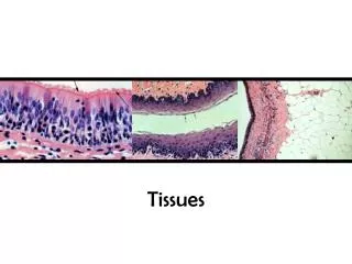

Tissues
E N D
Presentation Transcript
Hierarchy of organization • Atoms • Molecules • Cells • Tissues • Organs • Organ systems • Organism
Tissues are the fundamental units of organs and organ systems.
Tissues are the fundamental units of organs and organ systems.
Tissues are the fundamental units of organs and organ systems.
Tissues are the fundamental units of organs and organ systems.
Tissues • Histology is the study of tissues. • A tissue is a group of cells and intercellular materials that have a similar developmental origin, structure, and function. Loose connective tissue
What you should know about a tissue • Know the structure and be able to identify it from a slide. • Know the function. • Know location in the body it is likely to be found. • Know any special attributes of that tissue.
How to start identifying a tissue image. • Identify where the lumen (space) is in the image if you can. • Identify where the basement membrane is in the image is you can. The basement membrane is where the tissue stops. • Look at how many layers of cells there are. (1) • Look at the shape of the cells closest to the lumen. (long) • Look for other special features of the tissue. • Give the tissue a complete name. Image from: http://biology.clc.uc.edu/fankhauser/Labs/Bio_Lab113/Tissues/Human_Histology.html
An important thing to remember when looking under the microscope • To prepare tissue slides, scientists must slice through the tissue. They can slice in different ways. Cross section Longitudinal section
There are four basic tissue types. • Epithelial Tissue (Epithelium) • functions include protection, absorption, filtration, and secretion. • Connective Tissue • functions include protecting, supporting, binding together tissues, separating, storing energy, transporting materials. • Muscle Tissue • functions in the movement of the skeleton, pumping of the heart and the movement of food. • Nervous Tissue • send electrical signals through the body, thus forming a communication system through the body.
Characteristics of the epithelium • Cells fit closely together to form continuous sheets and are bound together at many points by cell junctions. • Cells have one free surface or edge. This apical surface is exposed to the body’s exterior or to a cavity (the lumen) • The lower cell surface rests on a basement membrane, a structureless material secreted by the cells. • These tissues are avascular, meaning that they have no blood supply and depend on diffusion from capillaries in the underlying connective tissue • If well nourished, they can regenerate easily.
Characteristics of the epithelium apical surface of cells near lumen basement membrane Image from: http://biology.clc.uc.edu/fankhauser/Labs/Bio_Lab113/Tissues/Human_Histology.html
Characteristics of the epithelium Number of Layers: Simple – single layer Stratified – multiple-layered Pseudostratified – only appears to be multi-layered
Characteristics of the epithelium Cell Shape:
Types of Epithelium • Simple Squamous Epithelium • Stratified Squamous Epithelium • Simple Cuboidal Epithelium • Stratified Cuboidal Epithelium • Simple Columnar Epithelium • Stratified Columnar Epithelium • Pseudostratified Columnar Epithelium • Transitional Epithelium
Epithelium is found everywhere. Simple squamous Epithelium lines alveoli in lungs. Stratified squamous Epithelium lines the esophagus. Simple cuboidal Epithelium Forms tubes in kidneys. Simple columnar Epithelium lines the intestine.
Simple squamous epithelium • Thin and leaky • Good for exchange of materials by diffusion • Blood vessels • Alveoli
Simple squamous epithelium These are the same tissue. Why do they look so different? Images from: http://biology.clc.uc.edu/fankhauser/Labs/Anatomy_&_Physiology/A&P201/Epithelium/Epithelial_Tissues.htm
Stratified squamous epithelium • Regenerates rapidly by division of cells at its attached surface • New cells move toward the free surface; older cells slough off • Suited for covering and lining surfaces subject to abrasion • 2 types – nonkeratinized and keratinized • keratin is a strong, fibrous protein
Stratified squamous epithelium Images from: http://biology.clc.uc.edu/fankhauser/Labs/Anatomy_&_Physiology/A&P201/Epithelium/Epithelial_Tissues.htm
Cuboidal epithelium • Simple Cuboidal epithelium • Secretion, absorption, protection • Ducts of many glands, covering of ovary, form kidney tubules • Stratified Cuboidal epithelium • Secretion, absorption • Lines ducts of sweat glands.
Cuboidal epithelium Kidney section Images from: http://biology.clc.uc.edu/fankhauser/Labs/Anatomy_&_Physiology/A&P201/Epithelium/Epithelial_Tissues.htm
Kidney tubules, glands, lining of terminal bronchioles, etc.
Simple columnar epithelium • Transportation, absorption, secretion, protection • Large surface area • Lines much of the digestive tract, gall bladder, and large ducts of glands • May have a brush border of microvilli • May be ciliated– uterus, small bronchi, and paranasal sinuses.
Simple columnar epithelium Brush border Cilia Images from: http://biology.clc.uc.edu/fankhauser/Labs/Anatomy_&_Physiology/A&P201/Epithelium/Epithelial_Tissues.htm
Stratified columnar epithelium • Surface cells are columnar • Secretion, absorption, protection • Some large excretory ducts, portions of the male urethra • No cilia • Not common
Pseudostratified columnar epithelium • Surface cells are columnar • Secretion, absorption, lubrication, protection, transportation • Lines most of trachea, primary bronchi, epididymis and ductus deferens, nasal cavity, male urethra, large excretory ducts. • Usually ciliated. • May contain goblet cells, which secrete mucous
Lines nasal cavity & sinuses, auditory tubes, trachea, bronchi
Transitional epithelium • Surface cells are dome-shaped when relaxes but flattened when streched. • Protection, distensible • Lines urinary tract
Transitional epithelium Distended bladder Image from: http://erl.pathology.iupui.edu/HISTO/GENER64.HTM
Glandular Epithelia - Endocrine & exocrine glands • Endocrine Glands - Release hormones into interstitial fluid; no ducts • Exocrine Glands - Produce secretions onto epithelial surfaces; through ducts Figure 4–6
Modes of Secretion • Are produced in Golgi apparatus • Are released by vesicles (exocytosis) • e.g.,sweat glands • Merocrine secretion • Apocrine secretion *Are produced in Golgi apparatus *Are released by shedding cytoplasm *e.g., mammary glands • Holocrine secretion • Are released by cells bursting, killing gland cells • Gland cells replaced by stem cells • e.g.,sebaceous gland Figure 4–6a
Connective Tissue • Characterized by the cells widely separated from each other in a matrix that is produced by the cells. • Tissue protects and supports. • Cell Matrix composed of two regions • Ground • Liquid (sol), Gel, Gum or solid • Fibers • Non-elastic (= white or Collagen) • Elastic (= yellow fibers) • Types of Connective tissue
Types of Connective Tissue • Loose (Areolar) Connective Tissue • Dense Connective Tissue • Adipose • Cartilage • Bone • Blood
Classification of Connective Tissues (3) • Connective tissue proper:connect and protect • Fluid connective tissues:transport • Supportive connective tissues:structural strength Connective Tissue Proper Categories • Loose connective tissue:more ground substance, less fibers e.g., fat (adipose tissue) • Dense connective tissue:more fibers, less ground substance e.g., tendons
8 Cell Types of Connective Tissue Proper • Macrophages-large, amoeba-like cells of the immune system: • eat pathogens and damaged cells, fixed macrophages stay in tissue, free macrophages migrate • Fibroblasts most abundant cell type-in all connective tissue proper & secrete proteins & hyaluronan (cellular cement) • Adipocytes-fat cells-each cell stores a single, large fat droplet • Mesenchymal Cells-stem cells that respond to injury or infection: differentiate into fibroblasts, macrophages, etc. • Melanocytes -synthesize and store the brown pigment melanin • Mast Cells -stimulate inflammation after injury or infection:release histamine and heparin • Basophils are mast cells carried by blood • Lymphocytes-specialized immune cells in lymphatic system: e.g.,plasma cells which produce antibodies • Microphages -phagocytic blood cells: respond to signals from macrophages and mast cells, e.g., neutrophils and eosinophils


