X-ray Crystallography-1
X-ray Crystallography-1. Crystal Properties, space groups Diffraction Bragg’s law, von Laue condition X-ray diffraction data collection. Reading: van Holde, Physical Biochemistry, Chapter 6; the two Watson & Crick papers Additional optional reading:

X-ray Crystallography-1
E N D
Presentation Transcript
X-ray Crystallography-1 • Crystal Properties, space groups • Diffraction • Bragg’s law, von Laue condition • X-ray diffraction data collection Reading: van Holde, Physical Biochemistry, Chapter 6; the two Watson & Crick papers Additional optional reading: Gale Rhodes, Crystallography Made Clear, sections of Chapters 1-4 Homework: (see next two pages), due Wednesday, Feb. 22 Remember: Pizza & Movie, Sun, Feb. 12, 6:00 pm Midterm 1: Monday, Feb. 27 Many slides adopted from Prof. W. Todd Lowther, Dept. of Biochemistry, Wake Forest University Additional slides adopted from Prof. Ernie Brown, formerly in the Dept. of Chemistry, Wake Forest University
Homework 4 (Chapter 6, X-ray diffraction), due Wednesday, Feb. 22 • If not stated otherwise, assume l = 0.154 nm (CuKa-radiation) • van Holde 6.1 • van Holde 6.2a (Hint: put one atom at x, y, z, the other atom at x+1/2, -y, -z) • van Holde 6.3 • NaCl crystals are crushed and the resulting microcrystalline powder is placed in the X-ray beam. A flat sheet of film is placed 6.0 cm from the sample and exposed. Ignoring the possibility of forbidden reflections (which is in fact the case with NaCl, because the lattice is centered), what would be the diameters and indices of the first two (innermost) rings on the photograph? NaCl is cubic with unit cell dimension a = 0.56nm. • You are working with a linear crystal of atoms, each spaced 6.28 nm apart. You adjust your x-ray emitter so that it emits 0.628 nm x-rays along the axis of the array. • As we did in class, using the reciprocal lattice and the sphere of reflection, draw the allowed S vectors (S = k - k0). Denote the direction of propagation of the incoming x-rays as the positive x-axis. (k0 is direction of incoming X-rays, S is scattering vector). • You place a 1 cm2spherical detector 1 cm from the sample, centered on the x-axis, on the opposite side of the emitter. Draw the pattern you expect to detect. Clearly mark the expected distances. • If you performed the experiment on a linear crystal with atoms spaced 0.1 nm apart, what pattern would you detect? Would you have the same pattern if your detector were 1 m2? What does this say about the resolution of your experiment? • … continued on next page
Homework 4 (Chapter 6, X-ray diffraction), due Wednesday, Feb. 22: • … continued • If not stated otherwise, assume l = 0.154 nm (CuKa-radiation) • van Holde 6.6 a-d (see Fig. 6.18) • van Holde 6.9 a-c (in c), real space means on film) • van Holde 6.9 d but: Sketch the fiber diffraction pattern expected for A-DNA (not Z-DNA).
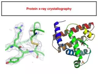
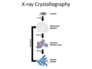
X-ray crystallography – in a nutshell • Protein is crystallized • X-Rays are scattered by electrons in molecule • Diffraction produces a pattern of spots on a film that must be mathematically deconstructed (Fourier transform) • Result is electron density (contour map) – need to know protein sequence and match it to density • coordinates of protein atoms put in protein data bank (pdb) download and view beautiful structures. • Currently there are about 80,000 structures in the pdb (2012). Check out protein data bank: (http://www.rcsb.org)
Fourier transform ? Phase Problem ? MIR MAD MR X-ray Crystallography – in a nutshell Reflections: h k l I σ(I) 0 0 2 3523.1 91.3 0 0 3 -1.4 2.8 0 0 4 306.5 9.6 0 0 5 -0.1 4.7 0 0 6 10378.4 179.8 . . . X-ray diffraction pattern Protein crystal Bragg’s law Fit molecules (protein) into electron density Electron density Electron density: r(x y z) = 1/V SSS |F(h k l)| exp[–2pi (hx + ky + lz) + ia(h k l)]
X-ray crystallography – in a nutshell End result!Fourier transform of diffraction spots electron density fit amino acid sequence DNA pieces Protein (Dimer of dimers)
Why determine the 3-D structure of your favorite protein or protein-ligand complex? • A picture is worth a thousand words. • Insight into structure-function relationships • Recognition and Specificity • Might identify a pocket lined with negatively-charged residues • Or positively charged surface – possibly for binding a negatively charged nucleic acid • Rossmann fold – binds nucleotides • Zinc finger – may bind DNA. • Aids in the design of future experiments • Rational drug design • Engineered proteins as therapeutics Chicken Fibrinogen S-Nitroso-Nitrosyl Human hemoglobin A
Visible light vs. X-raysWhy don’t we just use a special microscope to look at proteins? • Resolution is limited by wavelength. Resolution ~ l/2 • Visible light: 400-700 nm • X-rays: 0.7-1.5 Å (0.07-0.15 nm) • But to get images need to focus light (radiation) with lenses. • It is very difficult to focus X-rays (Fresnel lenses, doesn’t really work for X-rays) there are no lenses for X-rays can see atoms directly. • Getting around the problem X-ray Crystallography • Defined beam • Regular structure of object (crystal) • Result – diffraction pattern (not a focused image).
The Electromagnetic Spectrum • Wavelength of the “radiation” needs to be smaller than object size. • Diffraction limit (separation of resolvable features): ~ l/2
Crystal formation • Start with supersaturated solution of protein • Slowly eliminate water from the protein • Add molecules that compete with the protein for water (3 types: salts, organic solvents, PEGs) • Trial and error • Most crystals ~50% solvent • Crystals may be very fragile
What are crystals? • Ordered 3D array of molecules held together by non-covalent interactions Unit Cell • Sometimes see electrostatic or “salt interactions” • Lattice network • Defined planes of atoms
What are crystals? • Solids that are exact repeats of a symmetric motif • In a crystal, the level at which there is no symmetry asymmetric unit. • Apply rotational or screw operators to construct lattice motif. • Lattice motif is translated in three dimensions to form crystal lattice. • The lattice points are connected to form the boxes unit cell. • The edges define a set of unit vector axes unit cell dimensions a, b, c. • Angles between axes:
What are crystals? • Cystal stack unit cells repeatedly without any spaces between cells • Unit cell has to be a parallelepiped with four edges to a face, six faces to a unit cell. • All unit cells within a crystal are identical morphology of (macroscopic) crystal is defined by unit cell • There are only seven crystal systems (describing whole (macroscopic) crystal morphology): Triclinic, Monoclinic, Orthorhombic, Tetragonal, Trigonal, Hexagonal, Cubic (defined by length of unit vectors and angles). • There are only fourteen unique crystal lattices fourteen Bravais lattices. • P = primitive lattice point at corners of unit cell, F= face centered lattice point at all six faces, I = lattice point in center of unit cell, C = centered, lattice point on two opposing faces.
What are crystals?Bravais Lattices and Space Groups • 7 crystal systems • 14 Bravais lattice systems • Space group = • Lattice identifier + known symmetry relationships • Molecules within the crystal will most likely pack with symmetry
What are crystals?Bravais Lattices and Space Groups • What symmetry operations (e.g. rotation axes, (2-, 3-, 4-, 6- fold axis, mirrors, inversion axes …, at corner, at face, … (see Table 1.4) can be applied to the unit cell (inside crystal)? This defines the 32point groups of the unit cell. • The combination of the 32 symmetry types (point groups) with the 14 Bravais lattice, yields 230 distinct space groups. • In biological molecules, there are really only 65 relevant space groups (no inversion axes or mirrors allowed, because they turn L-amino acids into D-amino acids. • The space group specifies the lattice type (Bravais lattices, outside crystal morphology) and the symmetry of the unit cell (inside). • Different crystals that have identical unit cell lengths and angles and are in the same space group are isomorphous.
Examples of Symmetry • Rotations • 2-folds (dyad symbol) • 3-folds (triangle) • 4-folds (square) • 6-folds (hexagon) • Rotations can be combined • Translations • -moved along fractions of the unit cell • -see P21 example
(adapted from Bernhard Rupp, University of California-LLNL) http://www-structure.llnl.gov/Xray/tutorial/Crystal_sym.htm Examples Two-fold axis protein Bovine Pancreatic Trypsin InhibitorP 212121 (Primitive, orthorhombic unit cell with a two-fold screw axes along each unit cell vector)
What are crystals? Cell Edges, Angles, and Planes • Cell edges: a, b, c • Cell angles: a, b, g • (100), (001), (010) planes define the unit cell
What are crystals? Examples of 2-D Diagonal Planes, Miller planes, Miller indices • Diagonals through the unit cell denoted by how they cross-section an axis • e.g. 1/2 = 2, 1/3 = 3, 1/4 = 4, etc. • e. g.: (230) plane has intercepts at 1/2x and 1/3y • (-230) plane has intercepts at -1/2x and 1/3 y (slanted in other direction)
(230) planes b/3 a/2 (100) planes b a
What are crystals? Planes in 3-D and Negative Indices • Planes extend throughout crystal with different relationships to the origin: e.g. (234) • Negative indices tilt the plane the opposite direction: • NOTE that (210)=(-2-10)≠(-210). “-” sign usually put as a bar above the number
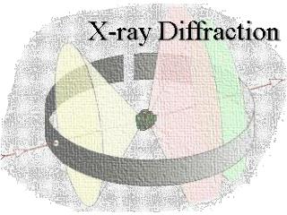
Theory of X-ray diffraction • Treat X-Rays as waves (CuKal ~ 0.154 nm). • Scattering: ability of an object to change the direction of a wave. • If two objects (A and B) are hit by a wave they act as a point source of a new wave with same wavelength and velocity (Huygens’ principle) • Diffraction: Those two waves interfere with each other. • destructive and constructive interference. • observe where maxima and minima are on screen. • get position of A and B Constructive interference: Destructive interference:
d Fig. 6.10 Bragg’s law: What is the relationship between diffraction angle 2q and unit cell dimensions? n… integer, l wavelength of X-ray Bragg’s law (simple model of crystal, but it works!)Crystal is made up of crystal planes (the Miller planes we just discussed). Assume a one-dimensional crystal: Geometric construction in class Reciprocal relationship between the Bragg angle q and the spacing, d, between the lattice planes. By measuring q, we can use Bragg’s law to determine dimension of unit cell!
In three dimensions (pp 263-265): 2q Each cone (h=1, -1, 2, -2 …) von Laue condition for diffractionNow we’ll move on to a three-dimensional crystal. Lattice still consists of only planes, but now we have a three-dimensional grid (still just dots, no internal structure, yet) h, k, l, are the Miller indices. Every discrete diffraction spot on a film has a particular Miller index. This are the same indices that describe the Miller planes. E. g. reflection (1,0,0) h=1, k=l=0; comes from (100) Miller plane 2q is the angle measured from the incoming X-ray beam
von Laue Condition for Diffraction “One-dimensional crystal” (horizontal planes) k = -2, -1, 0, 1, 2 “Three-dimensional crystal” (horizontal and vertical planes) (Horizontal and vertical diffraction cones, dots at intersections) h = 2 h = 1 h = 0 h = -1 h = -2 a c b
Determining the dimensions of the unit cell from the diffraction spots. k = 1, 2, 3 k = -1, -2, -3 h = 1, 2, 3,… h = -1, -2, -3,… Precession photograph of Tetragonal crystal of T4 lysozyme (X-ray aligned with third axis). Note: The spacing (angle q) is not affected by the number of molecules in a unit cell (more in a little bit).
Example A NaCl crystal is crushed and the resulting microcrystalline powder is placed in an 0.154 nm X-ray beam. A flat sheet of film is placed 6.0 cm from the sample. What is the diameter of the innermost ring on the photograph. NaCl is cubic with unit cell dimension 0.56 nm. (A powder gives diffraction rings instead of spots, because of the random orientation of the microcrystals in the powder.) Diffraction image: http://www.union.edu/PUBLIC/PHYDEPT/jonesc/images/Scientific/Powder%20diff%20Al.jpg
Determining the crystal symmetry from systematic absences Simple, conceptual example: P21 space group: Has a 2-fold screw axis along c-axis On 00l-axis only every other spot is observed. Each space group specifies its unique set of special conditions for observed and unobserved reflections along the principal and diagonal axes. (Can be looked up in tables). Sometimes it is very tricky to assign proper space group, especially for centered cells.
Translational symmetry elements and their extinctions.(Table 5.2 Jensen & Stout) Sometimes there is ambiguity, i.e. two space groups have same pattern
Is Bragg’s law still valid for two or more atoms in a unit cell? Conceptually: Jensen and Stout Two atoms in a unit cell (reflect) waves from their respective planes. The waves combine and form a resultant wave, that looks like it has been reflected from the original unit cell lattice plane. Diffraction spot is in the same place, but has different intensity (intensity of resultant wave). We assumed the electron density is in planes. In reality it is spread throughout the unit cell. Nevertheless, the derivation is still valid, since it can be shown that waves scattered from electron density not lying in a plane P, can be added to give a resultant as if reflected from the plane.
So far… By observing the spacing and pattern of reflections on the diffraction pattern, we can determine the lengths, and angles between each side of the unit cell, as well as the symmetry or space group in the unit cell. Still, how do we find out what’s inside the unit cell? (i.e. the interesting stuff, like proteins).