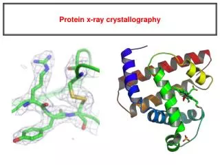
Introduction to protein x-ray crystallography
250 likes | 866 Vues
Introduction to protein x-ray crystallography. Electromagnetic waves. E- electromagnetic field strength A- amplitude w - angular velocity n - frequency l - wavelenght. E = A cos w t w = 2 pn. E = A cos ( a + w t ) a = 2pZ/l. a - phase. Wave as a vector. F=Acos a +iAsin a or

Introduction to protein x-ray crystallography
E N D
Presentation Transcript
Electromagnetic waves • E- electromagnetic field strength • A- amplitude • w- angular velocity • n- frequency • l - wavelenght E = A cos wt w = 2pn E = A cos (a+wt) a = 2pZ/l • a - phase
Wave as a vector F=Acosa+iAsina or F=exp(ia) Imaginary axis A A- wave amplitude a- wave phase a Real axis
What happens to electron when it is hit by x-rays? • The electron starts vibrating with the same frequency as the x-ray beam • As a result, secondary beams will be scattered in all directions Primary beam Secondary beams
Scattering from a molecule • Molecule is composed of many electrons • Each electron will scatter secondary radiation uppon exposure to x-rays • The scattered secondary beams will interact and cause interference • The scattering from a molecule is dependent on number of and distances between electrons • In other words, scattering from molecule is dependent on its structure • If we would know the amplitudes and phases of scattered molecule, we could calculate the structure of molecule... Primary beam
In practice... • Scattering from a single molecule is far too weak to be observed • If molecules are all oriented in the same way (like in crystal), the scattering from individual molecules will be multiplied in certain directions
Braggs law Scattered beams in phase, they add up Scattered beams not in phase, they cancel each other nl = 2d sinq http://www.eserc.stonybrook.edu/ProjectJava/Bragg/
Fourier transform • F(k)= f(x)e-2pikxdx • The electron density distribution of molecular structure and its produced diffraction pattern are fourier transforms respective to each other
The electron density equation • h,k,l – indices of reflections • xyz – coordinates • F – amplitude of reflections • a – phase of reflections • V- unit cell volume
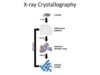
The Phase Problem • With detector you can measure only the intensity of reflections • The information about phases is lost – there is no such thing as “phase meter” • This means, you must obtain phase information in some other way • For small molecules (<100 atoms), direct methods exist. This means, that you can calculate phases from amplitudes without any extra information. • Proteins are far too big to use direct methods, so other tools are developed
Isomorphous replacement • By introducing heavy atoms in protein crystal (by soaking), the diffraction pattern can be altered • It is possible to determine positions of heavy atoms and from them the phases • One must use at least 2 different heavy atom soaks • Problems: • 1) Unit cell dimensions of crystal might change upon soaking • 2) Crystal might get destroyed upon soaking and not diffract at all • 3) Heavy atom ions might not bind in well defined places
Molecular replacement • Currently the most common technique • Applicable only if a similar structure already exists (at least 25% sequence identity) • The phases of known structure are combined with intensities of unknown • Before that, the known model has to be in silico placed in an artifical unit cell in the same orientation and translation from origin as in the structure of interest • For this, rotation and translation functions exist Problems: • May not work, if unknown structure is less than 30 % idendical to the known structure • Model bias – what’s that?
Phases unknown! Observed amplitudes Unknown structure FFT Cat Fourier cat Known structure Calculated amplitudes and phases Manx cat FFT Fourier Manx cat
Observed amplitudes, calculated phases FFT The tail becomes visible!
Be aware – this happens, if structures are not similar enough!! Duck Fourier duck Duck amplitudes + cat phases Looks like a cat!!
Model building • Fitting of protein sequence in the electron density • Easy in molecular replacement • More difficult if no initial model is available • Unambiquous if resolution is high enough (better than 3.0 Å) • Can be automated, if resolution is close to 2Å or better
Refinement • Automated improvement of the model, so it explains the observed data better • The phases get improved as well, so the electron density maps get better
Validation • Assesment of the final(?) model quality • How the geometry of amino acids look like? (Ramachandran plot) • Are non-covalently atoms far enough from each other? (no atom bumps) • Are residues “happy” in their environment? (hydrophobic in core, polar on surface) • Are the hydrogen donors/acceptors satisfied?
Depositing • Depositing of structure in PDB is required for the paper to be accepted in most journals • It is a good idea to deposit the diffraction data as well – this will prove that your structure actually has something to do with observed electron density