Download
1 / 41
410 likes | 609 Vues
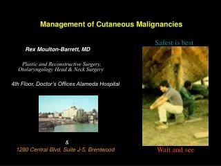
Management of Cutaneous Malignancies. Safest is best. Rex Moulton-Barrett, MD Plastic and Reconstructive Surgery, Otolaryngology Head & Neck Surgery 4th Floor, Doctor ’ s Offices Alameda Hospital & 1280 Central Blvd, Suite J-5, Brentwood.

E N D
Management of Cutaneous Malignancies Safest is best Rex Moulton-Barrett, MD Plastic and Reconstructive Surgery, Otolaryngology Head & Neck Surgery 4th Floor, Doctor’s Offices Alameda Hospital & 1280 Central Blvd, Suite J-5, Brentwood Versus: wait and see
The 8 Aspects of Plastic Surgery • Congenital: clefts, nevi, vascular tumors ear reconstruction, hand anomalies • Hand: nerve compression, tumors/soft tissue, trauma • Burn Reconstruction • General Reconstruction: truck, abdomen, lower limb • Breast: reduction, reconstruction • Cosmetic • Head and Neck: resection and reconstructivesurgery • Skin cancer: excision and reconstruction
Tumors In Question • Basal Cell • Squamous Cell • Melanoma • The differential diagnosis: non-pigmented benign pigmented benign non-pigmented pre-malignant pigmented pre-malignant soft tissue tumors metastatic lesions
Skin Cancer • Basal (75%) > Squamous (25%) > melanoma except organ transplant opposite ratio SCCA 20-65 times more common • 50% with basal or squamous will develop the other in 5 years • Intense > prolonged sun exposure: UVB>A, SPF 15, < 20 yrs age • Genetic predisposition: more pigment is protective
Common Non-pigmented Benign Lesions • Seborrheic Keratoses • Syringomas • Xantheloma Palpebrum • Premalignant Actinic Keratoses
Common non-pigmented benign lesions • Syringomas: peri-ocular , small, fleshy and nodular
Common non-pigmented benign lesions • Xantheloma Palpebrum: periocular, drop like & semi-cheezy rarely associated with hyperlipidaemia ie planar xanthomatadysbetalipoproteinemia or hypercholesterolemia
Common non-pigmented benign lesions • Trichoepitheliomas: periocular, drop like
Common non-pigmented benign lesions • Milia: periocular, drop like & semi-cheezy
Common non-pigmented Benign/pre-malignant lesions • Actinic Keratoses: 20% SCCA, dry, crusty • really pre-malignant
Pigmented Benign Lesions • Blue Nevus • Pigmented Seborrheic Keratosis • Giant Nevus
Pigmented benign lesions • Blue Nevus: intradermal and subcutaneous not pre-malignant
Pigmented benign lesions • Pigmented Seborrheic Keratosis: waxy, soft can rub off a little
Non-Pigmented Pre-malignant Lesions • Bowen’s Disease: red scaly patch of Squamous Cell Carcinoma in situ
Pigmented Benign & Pre-malignant Lesions • Giant Nevus: 1-2 % population Risk of developing melanoma related to size: > 20cm diameter adult > 2 palm size / body 5-20 % by 10, peak at 3-5 yrs > 1 palm size / face 5-20 % by 10, peak at 3-5 yrs
Role for topical anti-mitotic agents 5-fluorouracil imiquimod 5% cream ( Aldara ) aminolevulinic acid photodynamic therapy
Role for topical anti-mitotic agents 0.5% 5-fluorouracil ( 5gram $100 ) effective for actinic keratoses small in-situ lesions: BCCA not for invasive small = electrodesiccation/curettage or excision RNA analogue precursor & progressive DNA labelling Contraindicated in pregnancy: teratogenic VSD’s
Role for Topical anti-mitotic agents Imiquimod 5% cream ( Aldara ) Immunomodulator: activates monocytes,macrophages, Langerhan’s cells, T cell infiltrates, cytokines: interferons, interlekins, TNF effective for: actinic keratoses superficial basal cell carcinoma* probably no role for squamous cell ca* frequency related reactions are common *3 nights/ week for 6 weeks: 73% clearance rate at 12 weeks: higher clearance rates *
Actinic Cheilitis (AC) Smith et al, 2002: J AM Acad Dermatol 47(4):497-501 • 15 pts with biopsy proven AC • 3 x weekly for 4-6 weeks • 4 weeks later all lesions cleared • Specific Side effects continued in some cases throughout therapy: • pain, redness, swelling, ulceration
Role for topical anti-mitotic agents Aminolevulinic acid photodynamic therapy Levulan Kerastick 20% solution 17 minute blue light exposures 69% failure for superficial SCCA at 8 months 44% failure for superficial BCCA at 8 months Fink-Puches, et al, 1998 Arch Dermatol 134, 821-826. Category C : unknown side-effects pregnancy or breast feeding Not if porphyria Not if taking: oral hypoglycemic agents, sulpha, grseofulvin, phenothiazines, doxycycline, HCTZ diuretics
Basal Cell Carcinoma incisional biopsy • Basal Cell: elliptical wedge is better than shave punch biopsies work well if: adequate in width and depth preferably not from center nodular superficial ulcerated pigmented morpheiform
Basal cell carcinoma excisional biopsy • 1 high power field under frozen section/ Moh’s surgery • 3-5 mm margin from the clinical edge: rolled to flat
Squamous cell Biopsy • Squamous Cell:elliptical wedge: from periphery towards center better than shave 6-10 mm margin if excisional biopsy
Excisional Biopsy • Melanoma: closest margin to remove the lesion, do not shave, or wedge may use punch if completely excise • Sarcomas: closest margin to remove the lesion • Adnexal : closest margin to remove the lesion • Metastatic: closest margin to remove the lesion
Excisional Biopsy • Melanoma: closest margin to remove* the lesion, do not shave, or wedge may use punch if completely excise Superficial spreading Lentigo maligna * nodular amelanotic subungal Acral lentinous
Management of Melanoma • <0.75 mm deep: 1cm margin • 0.75cm - 1.25mm deep: 1cm margin & ? sentinel node • 1.25 mm-4mm deep: 1-2cm margin & sentinel node biopsy • >4mm deep: 1cm margin and use of lymphadenectomy unproven
S/P Shave of Melanoma • 2 schools of thought • Excisional biopsy and based on depth decide on size of margin using same parameters • Excise based at least the depth of the shave ie 1-2 cm margin, when in doubt take larger margin
Excisional Biopsy • Kaposi’s Sarcomas: closest margin to remove* HIV with CD4 <200/mm3 *
Excisional Biopsy • Adnexal/appendage: ductal or non-ductal closest margin to remove hamartoma hidrocystoma mixed tumor
Excisional Biopsy • Metastatic: closest margin to remove the lesion* melanoma breast adenocarcinoma*
Moh’s Surgery • Microscopic margin is preferable to macroscopic margin ie face in the ‘H zone’ reduced visible scar may reduce incidence of false negative margin • Recurrent lesions: depth and width defined prior to closure • Availability of service
Dangerous Problems • Midline Lesions Intranasal: glioma ( 15% CNS communication ) or encephalocele ( 100% commun ) Forehead: dermoid( 15% crista galli communication), encephalocele gliomas ( not lateral brow dermoid- no communication ) • Back: myelocele, meningomyelocele occiput and neck: encephalocele myelocele meningomyocele
Difficult Problems Problems • Zygomatic Arch to Angle of the Mandible Parotid tumors Lymph nodes: atypical TB inflammatory-children, metastatic node if > 1.5cm adult Branchial Cleft Cysts ( < 1-2 yrs: congenital, >2-15 yrs inflammatory, > 15 yrs neoplastic )
Difficult Problems • Merkel Cell Tumors • Subungal Pigmentation • Sebaceous Adenoma
Difficult Problems • Merkel Cell Tumors: biopsy if excisional will require later larger margin and possible lymph node dissection, may need metastatic work-up and tumor conference presentation
Difficult Problems • Subungal Pigmentation Acquired melanocytic nevus melanoma
Difficult Problems • Sebaceous Adenoma Warty lesion often in the scalp, can be salmon colored present at birth, hamartoma > 10 yrs : will form BCCA and 19% form syringocystadenoma
Surgical Principles • I. Have a plan:: H & P, iodine allergy, tetanus toxoid, irrigation, instruments, suture and needle, define the defect, method of closure, drain, dressing, antibiotics, post-op wound care and when to remove sutures. • II. Always have a lifeboat:: If closure does not work out have a second plan in mind, including placing a skin graft • III. Acknowledge cosmetic units: The face can be divided sub-units. Within each unit there are favorable skin tension lines ( with the pt. in the sitting position and animated ) which form at 90 degrees to the mimetic muscles. Scars are less conspicuous if they lie parallel to these natural creases. • IV. Control tension: Place all the tension below the epidermis or in the fascia. The majority of the blood supply is in the subdermal plexus ( SDP ): superficial to the subcutaneous fat. Undermine to distribute the tension over a wider area.
Clinical Examples A. 5mm chronic ulcer of the hand in a wrinkled 90 yr man a. important history duration, bleeding, numbness, other medical problems, medications, pacemaker, adenopathy, associated skin lesions b. important physical characteristics wipe lesion and look at shape: ulcer with irregular border, little pigmentation c. type of biopsy punch or wedge using lidocaine with epinephrine. single suture for hemostatis. d. definitive management path: SQ cell ca. If margin clear 6mm ellipse transversely ( using 3 to 1 rule length to width excision ) with local and tag margin for orientation. If final pathology margin positive or close ( < 5MM ) re-excise in OR with frozen section.
B. 3mm pigmented lesion on the lateral neck of a 33 yr old male Caucasian computer programmer • a. important history duration, bleeding, numbness, other medical problems, medications, pacemaker • b. important physical characteristics of lesion to make the diagnosis adenopathy, associated skin lesions, shape, elevation, border, pigmentation and texture: irregular border, irregular pigmentation, not raised and smooth • c. type of biopsy excise using 4mm punch full thickness into subcutaneous fat or elliptical excision (using 3 to 1 rule ) with lidocaine with epinephrine. 3 sutures for closure. • d. definitive management path: Malignant Melanoma depth 0.72 mm no evidence of intra-vascular invasion. ellipse 1cm margin favorable skin tension lines. check final pathology to confirm clear of tumor. present in tumor board.
C. 3cm chronic elevated lesion on the cheek of a 55 yr old lady • a. important history duration, bleeding, numbness, medical problems, medications, pacemaker • b. important physical characteristics of lesion to make the diagnosis adenopathy, associated skin lesions, wipe lesion, look at shape, ulcer with irregular border, little pigmentation c. type of biopsy biopsy punch or wedge using lidocaine with epinephrine. Do not use silver nitrate on face, use battery cautery, hyfercator or a single suture for hemostatis d. definitive management: in operating room with frozen section path: basal cell ca. Take >3mm margin and close wound along favorable tension lines with a local flap