Download

1 / 32
330 likes | 488 Vues
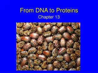
Chapter 12. From DNA to Proteins. Not a flashcard. The Size of DNA. If the DNA of one cell is stretched out, it makes a 9 ft. long string. There are about 7 trillion cells in the human body. The average human has enough DNA to go from the earth to the sun more than 400 times.

E N D
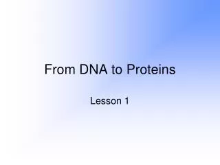
Chapter 12 From DNA to Proteins
Not a flashcard. The Size of DNA... If the DNA of one cell is stretched out, it makes a 9 ft. long string There are about 7 trillion cells in the human body The average human has enough DNA to go from the earth to the sun more than 400 times. (ruptured bacteria) That’s whole lot of information!
Not a flashcard. Why learn about DNA??? • YOU might be involved in needing… • A maternity/paternity test • Crime scene evidence analyzed • A child tested for genetic defects • Understanding antibiotic resistance for… • Humans • Crops • The future of medicine
Who discovered DNA? Edwin Chargaff 2 April 1953MOLECULAR STRUCTURE OF NUCLEIC ACIDS A Structure for Deoxyribose Nucleic Acid We wish to suggest a structure for the salt of deoxyribose nucleic acid (D.N.A.). This structure has novel features which are of considerable biological interest.A structure for nucleic acid has already been proposed by Pauling and Corey (1). They kindly made their manuscript available to us in advance of publication. Their model consists of three intertwined chains, with the phosphates near the fibre axis, and the bases on the outside. In our opinion, this structure is unsatisfactory for two reasons: (1) We believe that the material which gives the X-ray diagrams is the salt, not the free acid. Without the acidic hydrogen atoms it is not clear what forces would hold the structure together, especially as the negatively charged phosphates near the axis will repel each other. (2) Some of the van der Waals distances appear to be too small. Another three-chain structure has also been suggested by Fraser (in the press). In his model the phosphates are on the outside and the bases on the inside, linked together by hydrogen bonds. This structure as described is rather ill-defined, and for this reason we shall not comment on it. We wish to put forward a radically different structure for the salt of deoxyribose nucleic acid. This structure has two helical chains each coiled round the same axis (see diagram). We have made the usual chemical assumptions, namely, that each chain consists of phosphate diester groups joining ß-D-deoxyribofuranose residues with 3',5' linkages. The two chains (but not their bases) are related by a dyad perpendicular to the fibre axis. Both chains follow right- handed helices, but owing to the dyad the sequences of the atoms in the two chains run in opposite directions. Each chain loosely resembles Furberg's2 model No. 1; that is, the bases are on the inside of the helix and the phosphates on the outside. The configuration of the sugar and the atoms near it is close to Furberg's 'standard configuration', the sugar being roughly perpendicular to the attached base. There is a residue on each every 3.4 A. in the z-direction. We have assumed an angle of 36° between adjacent residues in the same chain, so that the structure repeats after 10 residues on each chain, that is, after 34 A. The distance of a phosphorus atom from the fibre axis is 10 A. As the phosphates are on the outside, cations have easy access to them. The structure is an open one, and its water content is rather high. At lower water contents we would expect the bases to tilt so that the structure could become more compact. The novel feature of the structure is the manner in which the two chains are held together by the purine and pyrimidine bases. The planes of the bases are perpendicular to the fibre axis. The are joined together in pairs, a single base from the other chain, so that the two lie side by side with identical z-co-ordinates. One of the pair must be a purine and the other a pyrimidine for bonding to occur. The hydrogen bonds are made as follows : purine position 1 to pyrimidine position 1 ; purine position 6 to pyrimidine position 6. If it is assumed that the bases only occur in the structure in the most plausible tautomeric forms (that is, with the keto rather than the enol configurations) it is found that only specific pairs of bases can bond together. These pairs are : adenine (purine) with thymine (pyrimidine), and guanine (purine) with cytosine (pyrimidine). In other words, if an adenine forms one member of a pair, on either chain, then on these assumptions the other member must be thymine ; similarly for guanine and cytosine. The sequence of bases on a single chain does not appear to be restricted in any way. However, if only specific pairs of bases can be formed, it follows that if the sequence of bases on one chain is given, then the sequence on the other chain is automatically determined. It has been found experimentally (3,4) that the ratio of the amounts of adenine to thymine, and the ration of guanine to cytosine, are always bery close to unity for deoxyribose nucleic acid. It is probably impossible to build this structure with a ribose sugar in place of the deoxyribose, as the extra oxygen atom would make too close a van der Waals contact. The previously published X-ray data (5,6) on deoxyribose nucleic acid are insufficient for a rigorous test of our structure. So far as we can tell, it is roughly compatible with the experimental data, but it must be regarded as unproved until it has been checked against more exact results. Some of these are given in the following communications. We were not aware of the details of the results presented there when we devised our structure, which rests mainly though not entirely on published experimental data and stereochemical arguments. It has not escaped our notice that the specific pairing we have postulated immediately suggests a possible copying mechanism for the genetic material. Full details of the structure, including the conditions assumed in building it, together with a set of co-ordinates for the atoms, will be published elsewhere. We are much indebted to Dr. Jerry Donohue for constant advice and criticism, especially on interatomic distances. We have also been stimulated by a knowledge of the general nature of the unpublished experimental results and ideas of Dr. M. H. F. Wilkins, Dr. R. E. Franklin and their co-workers at King's College, London. One of us (J. D. W.) has been aided by a fellowship from the National Foundation for Infantile Paralysis. J. D. WATSON F. H. C. CRICK Medical Research Council Unit for the Study of Molecular Structure of Biological Systems, Cavendish Laboratory, Cambridge. April 2. 1. Pauling, L., and Corey, R. B., Nature, 171, 346 (1953); Proc. U.S. Nat. Acad. Sci., 39, 84 (1953). 2. Furberg, S., Acta Chem. Scand., 6, 634 (1952). 3. Chargaff, E., for references see Zamenhof, S., Brawerman, G., and Chargaff, E., Biochim. et Biophys. Acta, 9, 402 (1952). 4. Wyatt, G. R., J. Gen. Physiol., 36, 201 (1952). 5. Astbury, W. T., Symp. Soc. Exp. Biol. 1, Nucleic Acid, 66 (Camb. Univ. Press, 1947). 6. Wilkins, M. H. F., and Randall, J. T., Biochim. et Biophys. Acta, 10, 192 (1953). Linus Pauling James Watson & Francis Crick April 2, 1953 Maurice Wilkins Rosalind Franklin
Not a flashcard. History of DNA • James Watson & Francis Crick (1953)- • First to discover DNA structure. • Edwin Chargaff (1952)- • Discovered the amount of adenine equals the amount of thymine AND the amount of cytosine equals the amount of guanine. • Rosalind Franklin (1952)- • Photographed DNA using X-ray crystallography.
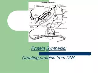
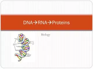
Protein Synthesis The steps from DNA to Proteins. DNA mRNA Amino Acids Proteins
DNA Begin here! • Deoxyribonucleic acid • Deoxyribose = ribose sugar missing 1 oxygen • Double stranded • Made of 3 parts: • Deoxyribose sugar • Phosphate group • Nitrogen base (ATCG) • Found mostly inside the nucleus • Too big to get out!
mRNA • Messenger RNA (RNA= ribonucleic acid) • Single stranded • Made of 3 parts: • Ribose sugar • Phosphate group • Nitrogen base (AUCG) • Copied from DNA • *this process is called Transcription!!! • Travels from the nucleus to the cytoplasm • DNA is too large to do this.
Amino Acids • A ribosome uses the directions from mRNA to pair the correct amino acid • Amino acids are on tRNA’s • This happens in the cytoplasm. A.A. A.A. tRNA
Protein Stop here! • A chain of amino acids is a protein. • Different proteins are made by: • Using different amino acids • Using longer or shorter chains of amino acids. • Translation- the process of making a protein. Proteins are determined by the chain of amino acids that make them up!
Vocabulary • Codon- 3 bases found on mRNA • Codes for an amino acid. • Example: CCG codes for glycine • Start & Stop codons control beginning and ending • Anti-codon- 3 bases found on tRNA • Example: GGC has glycine attached • tRNA- transfer RNA. Has an amino acid attached. Brings it to mRNA. More vocabulary!
More Vocabulary • Gene- a sequence of nucleotides that controls the production of 1 specific protein. • Replication- the copying of DNA. • Happens in the nucleus. • Very simple: DNA unzips and new bases attach. • MUST be copied to make new cells!
Not a flashcard. Recap… DNA mRNA Amino Acids Proteins Transcription- • From DNA to mRNA • Inside the nucleus Translation- • From mRNA to protein • Outside the nucleus (in the cytoplasm)
Translation The process of making proteins
Not a flashcard. Transcription anticodon Translation
Not a flashcard. Another View
Not a flashcard. And 1 more…
Mutation • Any change in the DNA sequence. • Can be helpful or harmful. What happens if you copy a phone number incorrectly? 875-2245 and 875-2235 are NOT the same or necessarily even close!
Mutagens • Anything that causes a mutation. • Chemicals • X-rays • UV light • Radioactivity
5 Types of Mutations • Point mutation • Framshift mutation • Inversion • Translocation • Nondisjunction
1. Point Mutation • ONE BASE is changed. • This only affects ONE amino acid!!! • Example: • The fat cat ran and ran. • The fat rat ran and ran.
2. Frameshift Mutation • One base is added (insertion) or deleted (deletion). • Affects that amino acid AND all thereafter! • Example: • The fat cat ran and ran. • The fat gca tra nan dra n (insertion) • The atc atr ana ndr an (deletion)
3. Inversion • Part of a chromosome breaks off and reattaches backwards.
4. Translocation • Part of a chromosome breaks off and reattaches to another chromosome!!!
5. Nondisjunction • Chromosomes don’t separate during meiosis. • Will cause: • Monosomy- 1 chromosome pair • Trisomy- 3 chromosome pairs Normal Normal
Not a flashcard. Normal division Non-disjunction
How can organisms be different from each other if they are made up of the same molecules? • Only 1 reason: different order of nucleotides.
Not a flashcard. Codon Chart Can you find…? GAU UAA GUG UGG AUG AGU aspartate stop codon valine tryptophan start codon serine
Not a flashcard. There are only 20 Amino Acids
Not a flashcard. Sickle Cell Revisited
Not a flashcard. It’s all connected!!!