Download

1 / 25
250 likes | 727 Vues
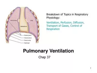
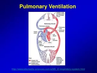
Pulmonary Ventilation. Pulmonary Structure and Function. Pulmonary Ventilation – Process by which ambient air is moved into and exchanges with air in the lungs …Different from oxygen consumption. Figure 12.1. Pulmonary Structure and Function.

E N D
Pulmonary Structure and Function Pulmonary Ventilation – Process by which ambient air is moved into and exchanges with air in the lungs …Different from oxygen consumption Figure 12.1
Pulmonary Structure and Function Gas exchange(O2 and CO2) occurs in the alveoli O2 transfers from alveolus to capillary blood CO2 transfers from capillary blood to alveolus Each minute at rest 250 ml of O2 and 200 ml of CO2 diffuse in opposite directions Fig 12.1
Pulmonary Structure and Function Lungs contain 600 million alveoli Extremely thin-walled sacs (0.3 mm thick) Lie side by side with thin walled capillaries Alveoli receive largest blood supply of any organ in the body Fig 12.1
Pulmonary Structure and Function Lungs provide the gas exchange surface separating blood from alveolar gases Lungs – Extremely large surface areafor gas exchange Figure 12.2 Adult lung weighs 1 kg and hold 4-6 L of air
Pulmonary Structure and Function Pulmonary ventilation functions primarily to maintain a constant and favorable concentration of O2 and CO2 in the alveoli during rest and exercise Adequate pulmonary ventilation ensures complete gas exchange before blood leaves lungs for transport to tissues
Pulmonary Structure and Function Breathing mechanics Fig 12.3 Inspiration – diaphragm descends, ribs are raised, volume increases, intrapulmonic pressure decreases, air rushes in (chest cavity size increases) Contributing muscles: external intercostals, sternocleidomastoids, scalenes, spinal extensors
Pulmonary Structure and Function Breathing mechanics Fig 12.3 Expiration – Passive process diaphragm relaxes, ribs lower, volume decreases, intrapulmonic pressure increases, air rushes out (chest cavity size decreases) Contributing muscles: rectus abdominus, internal intercostals, posterior inferior serratus
Pulmonary Structure and Function Ventilatory System: Fig 12.4 Conducting Zone – Trachea to Bronchioles No alveoli Air transport, warming, humidification, particle filtration Anatomic "Dead" Space *Respiratory Zone – Bronchioles to Alveoli Surface area for gas exchange
Pulmonary Structure and Function Measuring Lung Volume: Lung volumes measurements (static or dynamic) can help identify potential obstructive or restrictive lung diseases Fig 12.6 Lung volumes vary with age, gender, body size, body composition and stature Water-sealed, volume displacement recording spirometer
Pulmonary Structure and Function Static Lung Volume Measurements: Provides record of ventilatory volume and breathing rate Fig 12.6 Tidal Volume (TV) – volume inspired or expired per breath (600 ml) Inspiratory Reserve Volume (IRV) – maximal inspiration at end of tidal inspiration (3000 ml) Expiratory Reserve Volume (ERV) – maximal expiration at end of tidal expiration (1200 ml) Force Vital Capacity (FVC) – maximal volume expired after maximal inspiration (TV+IRV+ERV; 4800 ml)
Pulmonary Structure and Function Static Lung Volume Measurements: Fig 12.6 Residual Lung Volume (RLV) – air volume remaining in lungs after maximal expiration (1200 ml) -allows uninterrupted gas exchange between blood and alveoli Functional Residual Capacity (FRC) – Volume in lungs after tidal expiration(ERV + RLV; 2400 ml) Total Lung Capacity (TLC) – volume in lungs after maximal inspiration (FVC + RLV; 6000 ml)
Pulmonary Structure and Function Dynamic Lung Volumes: Adequate pulmonary ventilation depends on ability to sustain high airflow levels (not air movement in single breath) • Dynamic Ventilation depends on: • FVC (“stroke volume” of the lungs) • Breathing rate High airflow levels (velocity) depends on lung compliance (ability to stretch or expand): “loose” (high compliance) – emphysema, asthma “stiff” (low compliance) – fibrosis
Pulmonary Structure and Function Too Much Elastic Recoil (stiff) No Elastic Recoil (loose) Dynamic Lung Volumes Fig 12.8 Forced Expiratory Volume (FEV) to FVC ratio: FVC measured over 1 s (FEV1.0) – measures pulmonary airflow capacity, or overall resistance to air movement upstream in the lungs (normal value = 80-85% of FVC)
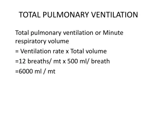
Pulmonary Structure and Function • 3. Minute Ventilation (VE): • Volume of air moved in and out of respiratory tract per minute • VE=Breathing Rate (BR) x TV • At rest VE = 12 breaths/min x 0.5 L/breath • VE = 6 Lmin • Exercise VE = 30 x 2.5 VE=50 x 3.5 • VE = 75 Lmin VE=150 Lmin Moderate Vigorous
Pulmonary Structure and Function • Dynamic Lung Volumes: • Measurements of dynamic lung function can indicate the severity of obstructive or restrictive lung diseases • FEV/FVC - Normal or increased for restrictive lung disease (80% or greater) • FEV/FVC - <70% indicates obstructive lung disease
Pulmonary Structure and Function Aging and lifestyle affect lung volumes and pulmonary function Aging: Decreased lung compliance FEV1.0 and FVC decrease after age 20 Diffusion capacity decreases Partial Pressure of O2 decreases Diaphragm muscles weakens by ~25%
Pulmonary Structure and Function Dynamic Lung Volumes: Provide no information about aerobic fitness: No difference in healthy vs olympic athletes Not predictive of track or marathon performance, distance running
Pulmonary Structure and Function • Dynamic Lung Volumes: • Important part of standard medical/health examination for “at risk” exercisers (smokers, asthmatics)
Pulmonary Structure and Function How do we ensure sufficient air reaches the alveoli during exercise? By increasing rate and depth of breathing - increases alveolar ventilation Fig 12.10 • TV increases at start of moderate exercise • As intensity increases, TV plateaus at 60% of FVC • Breathing rate provides alveolar ventilation at higher exercise intensities
Pulmonary Structure and Function Definitions: Hyperventilation – increase in pulmonary ventilation that exceeds the O2 needs of metabolism (“overbreathing”) Unloads CO2 excessively (which constricts arteries with less O2 to brain) Can lead to unconsciousness.
Pulmonary Structure and Function Definitions: Dyspnea – shortness of breath or subjective distress in breathing (sense of inability to breathe) Occurs in physical exertion (novel exercisers), at altitude, or with obstructive or restrictive pulmonary disorders Result of elevated CO2 and H+ in blood from fatigue of poorly trained respiratory muscles (shallow, ineffective breathing)
Pulmonary Structure and Function Definitions: Valsalva Maneuver – Increases intrathoracic pressure that occurs when exhalation is forced against a closed glottis Results: Collapse of veins in thoracic region Impaired venous return Acute DROP in arterial blood pressure Decreased blood supply to brain "spots before the eyes" "fainting"
Pulmonary Structure and Function Definitions: Valsalva Maneuver Acute drop in blood pressure Blood pressure overshoot Fig 12.11
Pulmonary Structure and Function Valsalva Maneuver *Valsalva Maneuver does NOT cause the acute rise in blood pressure with resistance training Fig 12.11