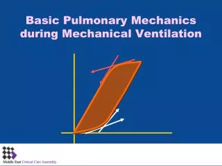
Advanced Pulmonary Mechanics during Mechanical Ventilation
1.06k likes | 2.13k Vues
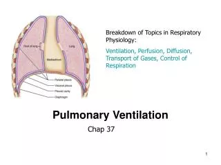
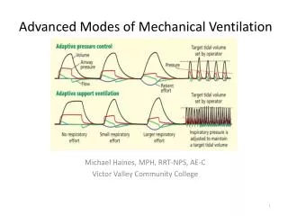
Advanced Pulmonary Mechanics during Mechanical Ventilation. Points of Discussion. Basics. Abnormalities. Equation of motion Dynamic Compliance Pressure-volume loop Flow-volume loop Work of breathing Lower and upper inflection points Hysterexis Intrathoracic Pressures. Air-leak

Advanced Pulmonary Mechanics during Mechanical Ventilation
E N D
Presentation Transcript
Points of Discussion Basics Abnormalities Equation of motion Dynamic Compliance Pressure-volume loop Flow-volume loop Work of breathing Lower and upper inflection points Hysterexis Intrathoracic Pressures Air-leak Air trapping Increased airway resistance Inadequate flow support Inadequate sensitivity Atelectasis Inadequate PEEP Airway obstruction Over-distension
Tube + Spring Model Resistive Forces Elastic Forces
Static and Dynamic Pressures Pressure PIP Flow-Resistive Pressure difference (Pres) Pplat Alveolar Distending (recoil) Pressure difference (Pdis) PEEP time
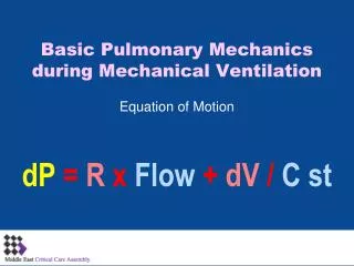
Resistive and Elastive Forces DYNAMIC CHARACTERISTICS: dP = dV / Cdyn RESISTANCE: dPresistive= R x Flow STATIC COMPLIANCE: dPdistensive = dV / Cst dP = dPresist. + dP dist. dP = R x Flow + dV / C st
Assessment of static P-V curveSuper-syringe method: Volume • Stepwise inflation from a big syringe with multiply occlusions at each volumes to record recoil pressure • Time consuming • Cumbersome to perform • Difficult to standardize • Patient must be paralysed, connected to a special equipment • Great risk of oxygen desaturation Pressure
Assessment of static P-V curveSlow Flow Single Inflation Method • Slow (5-10 lpm) inspiratory flow with large Vt and ZEEP • The inspiratory curve of the dynamic P-V loop closely approximates the static curve • The flow-resistive pressure component could be subtracted • Easy to perform, fast and relatively comfortable Servillo: AJRCCM 1997 Lu: AJRCCM 1999 Volume Static curve UPIflex inspiration LPIflex Pressure
Normal Compliance TLC VOLUME FRC FRC Negative Positive 0 DISTENDING PRESSURE FRC and PV Loop
Components of Pressure-Volume Loop VT Expiration Volume (mL) Inspiration PIP Paw (cm H2O)
Pressure-Volume Loop(Type of Breath) E E Vol (ml) E I I I Paw (cm H2O) Spontaneous Controlled Assisted I: Inspiration E: Expiration
VT PEEP PIP PEEP and P-V Loop Volume (mL) Paw (cm H2O)
Upper Inflection Point Lower Inflection Point Inflection Points • Upper Inflection Point: Represents pressure resulting in regional overdistension • Lower Inflection Point: Represents minimal pressure for adequate alveolar recruitment Volume (mL) Pressure (cm H2O)
Normal Patient Decreased Compliance Volume(ml) Pressure (cm H2O)
Lung Compliance Changes and the P-V Loop Volume Targeted Ventilation Preset VT Increased Normal Decreased Volume (mL) Paw (cm H2O) PIP levels
Lung Compliance Changes and the P-V Loop VT levels Increased Normal Pressure Targeted Ventilation Decreased Volume (mL) Preset PIP Paw (cm H2O)
Hysteresis Volume (ml) Normal Hysteresis Abnormal Hysteresis Pressure (cm H2O)
Flow-Volume Loop Inspiration PIFR Volume (ml) FRC VT Flow (L/min) PEFR Expiration
Positive Pressure Ventilation: The Equation of Motion • In a passive subject, airway pressure represents the entire pressure (P) applied across the respiratory system. • The work required to deliver a tidal breath (Wb) = tidal volume (VT) x airway pressure • The pressure (P) associated with the delivery of a tidal breath is defined by the simplified equation of motion of the respiratory system (lungs & chest wall): P = VT/CR+ VT/Ti x RR + PEEP total P elastic P resistive P elastic Where CR = compliance of the respiratory system, Ti = inspiratory time and VT/Ti = Flow, RR = resistance of the respiratory system and PEEP total = the alveolar pressure at the end of expiration = external PEEP + auto (or intrinsic) PEEP, if any. Auto PEEP = PEEP total – P extrinsic (PEEP dialed in the ventilator) adds to the inspiratory pressure one needs to generate a tidal breath.
Work of Breathing Volume (ml) B A: Resistive Work B: Elastic Work A Pressure (cm H2O)
Work of Breathing • WOB is a major source of caloric expenditure and oxygen consumption • Appr. 70% to overcome elastic forces, 30% flow-resistive work • Patient work is a one of the most sensitive indicator of ventilator dependency • Comparison of Ventilator and Patient work is a useful indicator during weaning process • WOB may be altered by changes in compliance, resistance, patient effort, level of support, PEEP, improper Ti, demand system sensitivity, mode setting • Elevated WOB may contraindicate the weaning process
WOB =∫0ti P x Vdt Elasic work: ABCA Resistive work Inspiratory: ADCA Expiratory: ACEA WOB Measurements V B C E D P A
Work of Breathing Measurements WOB =∫0ti P x Vdt • Paw: Ventilator Work: The physical force required to move gas into the lung, represents the total work of the resp. system (patient + ventilator) • Peso: Patient Work: done by respiratory muscles, represents the pulmonary work of breathing • Paw-Ptr: Imposed Work by the Endotracheal tube
P-V Loop and WOB V Normal Compliance Increased Resistance Decreased Compliance Normal Resistance P V Normal Compliance Normal Resistance V P P
Work of Breathing • Work per breath is depicted as a pressure-volume area • Work per breath (Wbreath)= P x tidal volume(VT) • Wmin = wbreath x respiratory rate WEL = elastic work WR = resistive work Volume Volume Volume VT Pressure Pressure Pressure The total work of breathing can be partitioned between an elastic and resistive work. By analogy, the pressure needed to inflate a balloon through a straw varies; one needs to overcome the resistance of the straw and the elasticity of the balloon.
Intrinsic PEEP and Work of Breathing When present, intrinsic PEEP contributes to the work of breaking and can be offset by applying external PEEP. Volume VT VT Dynamic Hyperinflation FRC Pressure PEEPi PEEPi = intrinsic or auto PEEP; green triangle = tidal elastic work; red loop = flow resistive work; blue rectangle = work expended in offsetting intrinsic PEEP (an expiratory driver) during inflation
The Pressure and Work of Breathing can be Entirely Provided by the Ventilator (Passive Patient) Ventilator + + ₊ + + ₊
The Work of Breathing can be Shared Between the Ventilator and the Patient The ventilator generates positive pressure within the airway and the patient’s inspiratory muscles generate negative pressure in the pleural space. AC mode PAW patient machine PES time Paw = Airway pressure, Pes= esophageal pressure
Work of breath Resistive Work Elastic Work of Lung Pressure Elastic Work of Chest Paw Pes Work to inflate the chest wall Inflation Deflation Volume
Relationship Between the Set Pressure Support Level and the Patient’s Breathing Effort The changes in Pes (esophageal pressure) and in the diaphragmatic activity (EMG) associated with the increase in the level of mask pressure (Pmask = pressure support) indicate transfer of the work of breathing from the patient to the ventilator. Carrey et al. Chest. 1990;97:150.
Partitioning of the Workload Between the Ventilator and the Patient • How the work of breathing partitions between the patient and the ventilator • depends on: • Mode of ventilation (e.g., in assist control most of the work is usually done by the ventilator) • Patient effort and synchrony with the mode of ventilation • Specific settings of a given mode (e.g., level of pressure in PS and set rate in SIMV)
Respiratory Mechanics in ARF* • Reduced range of volume excursion: Low compliance • Flattering at low and high volumes: Lower and upper inflection points *Bigatello: Br J Anaest 1996 Volume NORMAL ARDS Pressure
Lung Protective Strategy • Set PEEP above the lower Pflex to keep the lung open and avoid alveolar collapse • Apply small Vt to minimize stretching forces • Set Pplat below the upper Pflex to avoid regional overdistension Volume Pressure
Abnormalities • Air-leak • Air trapping • Increased airway resistance • Inadequate flow support • Inadequate sensitivity • Atelectasis • Inadequate PEEP • Airway obstruction • Over-distension
Air Leak Volume (ml) Air Leak Pressure (cm H2O)
Air Leak Inspiration Flow (L/min) Volume (ml) Air Leak in mL Normal Abnormal Expiration
Air Leak Volume (mL) Time (sec)
Air Trapping Auto-PEEP Normal Patient Air Trapping Inspiration Time (sec) Flow (L/min) } Expiration
Air Trapping Inspiration Flow (L/min) Does not return to baseline Volume (ml) Normal Abnormal Expiration
Response to Bronchodilator After Before Time (sec) Flow (L/min) Long TE PEFR Shorter TE Higher PEFR
Increased Airway Resistance Inspiration Flow (L/min) Volume (ml) Normal Abnormal “Scooped out” pattern Decreased PEFR Expiration
Increased Raw Higher PTA Vol (mL) Normal Slope Lower Slope Pressure (cm H2O)
Airway Secretions/Water in the Circuit Inspiration Flow (L/min) Volume (ml) Normal Abnormal Expiration
After Suction Before Suction Airway Obstruction F F V V
PEEP: 3 cmH2O PEEP: 8 cmH2O Optimising PEEP V V P P
Inadequate Sensitivity Volume (mL) Paw (cm H2O) Increased WOB
Replaced FRC Lost FRC Atelectasis V V P P
Overdistension With little or no change in VT Normal Abnormal Volume (ml) Pressure (cm H2O) Paw rises
Overdistension • Overdistension occurs when the volume limit of some components of the lung has been exceeded • Abrupt decrease in compliance at the termination of inspiration • Results in a terminal “Beaking” of the P/V Loop Volume Pressure
Overdistension Index Volume C20 Cdyn Pressure 0.8 Pmax Pmax