Download
1 / 84
860 likes | 1.1k Vues
Inflammatory Bowel Disease. Dr. Mohammad Shaikhani CABM, FRCP. Pretest 1:. What % of IBD is regarded to be intederminate : A.90% B. 70% C.10%. D.25%. E.0%. Pretest 2:. Appendisectomy in crease the risk of: A.UC B. CD C. Both. D. Neither. Pretest 3:. Smoking:

E N D
Inflammatory Bowel Disease Dr. Mohammad Shaikhani CABM, FRCP
Pretest 1: What % of IBD is regarded to be intederminate: A.90% B. 70% C.10%. D.25%. E.0%.
Pretest 2: Appendisectomy in crease the risk of: A.UC B. CD C. Both. D. Neither.
Pretest 3: Smoking: A. Increase the risk of UC. B. Protect against UC. C. Protect against CD. D. ALL. E. None..
Pretest 4: Proctitis in UC leads to: A. Constipation > diarrhea. B. Diarrhea> constipation. C. Both. D. Neither.
Pretest 5: CD can cause the following except: A. GOO. B. Mouth ulcer. C. Perianal fistulas. D. diverticuli. E. RIF mass
Pretest 6: CD can cause the following except: A. GOO. B. Mouth ulcer. C. Perianal fistulas. D. diverticuli. E. RIF mass
Pretest 7: Granulomas, characteristic of CD, occur in: A. Nearly all. B. Nearly none. C. Around 1/3. D. Most of them. E. None of the above.
Pretest 8: Primary sclerosingcholangitis: A. Occurs in CD > UC. B. Occurs in most patients with UC. C. Most patients with PSC have UC. D. All of the above. E. None of the above.
Pretest 9: IBD with Primary sclerosingcholangitis have increases risk of: A. CRC. B. Cholangiocarcinoma. C. Both. D. Neither.
Pretest 10: The most serious acute complications of IBD is: A. Pyodermagangrenosa. B. Toxic megacolon. C. PSC. D. EN. E. Peripheral arthritis.
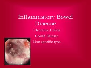
Introduction • IBD: an idiopathic chronic inflammatory disease of the GIT consistsing of 2 distinct clinical entities: ulcerative colitis & Crohn disease (regional enteritis),both cause macroscopic inflammation. • Microscopic colitis is less common& does not cause significant macroscopic abnormalities. • The pathogenesis not fully understood but likely involves a genetic predisposition& a dysregulated immunologic response to the local microenvironment of luminal bacteria. • Both are usually differentiated on the basis of differences in the distribution of pathology in the bowel & histopathologic appearance of the lesion. • 10% of patients cannot be shown to have either Crohn disease or ulcerative colitis ( indeterminate colitis).
Risk factors: The peak incidence :second, third& fourth decades of life A second peak in the seventh&eighth decades. There are no sex differences. Ashkenazi Jewish descent have a higher risk. A 5-10% risk for first-degree relatives of affected patients. Many candidate genes: 1.Established link between Crohn disease &variants of the CARD15 (also known as NOD2) gene, in only some patients & does not affect the risk for colitis. 2. IL-23 receptor affects the risk for both Crohn disease & colitis., as monoclonal antibody against the p40 subunit have demonstrated benefit in the treatment of Crohn disease.
Less common colitis forms are: • Microscopic colitis (collagenous& lynphocytic) • Others • Diversion colitis after clostomies. • Radiation colitis • Drug induced colitis • Infectious colitis • Ischemic colitis
Clinical features: UC • Generally present with bloody diarrhea with rectal urgency, discomfort& cramps. • They have profound tenesmus (feelings of urgency& incomplete evacuation), secondary to proctitis, this can cause constipation to be a more common manifestation than diarrhea; in such patients, determining the activity of the disease& treating it can be challenging. • UC extends proximally from the anal verge&can progress to pancolitis involving the cecum. • Fever is infrequent, but weight loss secondary to the inflammatory disease itself or to the chronic diarrhea is common. • Physical examination findings can range from mild lower abdominal tenderness to abdominal distention with rebound tenderness& hypoactive bowel sounds, suggestive of toxic megacolon.
Clinical features: Crohn disease More protean in its manifestations than ulcerative colitis, as the disease can affect any portion of the GIT& frequently has so-called “skip lesions” with areas of normal mucosa juxtaposed with severe inflammation. The transmural nature of the disease results in three distinct manifestations: inflammatory, fistulizing& fibrostenotic. Large-volume diarrhea can occur; diarrhea is associated with both small& large-bowel Crohn disease, whereas hematochezia is almost always a sign of colonic disease. The inflamed tissue causes a secretory diarrhea& a protein-losing enteropathy, steatorrhea from fat malabsorption (with patients who have ileal or ilealocolic disease frequently being vitamin B12- & vitamin D-deficient)& other types of malabsorption. Patients who have had their terminal ileum resected are also at risk for a choleretic diarrhea secondary to bile salt wasting.
Clinical features: Crohn disease Fistulae are abnormal connections between the bowel& adjacent organs. Abscesses may form& the fistula acts as a natural drainage mechanism, causing pus to emerge from the fistulae. The fistulae become symptomatic with drainage of fecal material around the anus (perianal fistulae), seepage of bowel contents through the skin (enterocutaneous fistulae), passage of feces through the vagina (rectovaginal fistulae)& pneumaturia or recurrent urinary tract infections (enterovesical fistulae). The intestinal inflammation may extend to adjacent musculature &result in neuromuscular sequelae; for example, a patient with Crohn disease&a new limp likely has a psoas muscle abscess.
Clinical features: Crohn disease Patients with intestinal strictures present with signs of obstruction: fever, abdominal distention, pain, nausea, vomiting. Strictures may be secondary to severe inflammation or to fibrosis of the bowel& can be relieved only by surgical resection. The most common site of strictures is in the terminal ileum where they result in partial or complete small-bowel obstruction. Patients with duodenal Crohn disease may develop gastric outlet obstruction. In patients with ileal disease, the abdominal examination commonly shows right lower quadrant tenderness; a phlegmonous mass may be present. A detailed anorectal examination is important in patients with suspected Crohn disease: the presence of skin tags suggests the diagnosis& the examination may also show fistulae.
Environmental Precipitants • Factors: • Early appendectomy (increase UC incidence) • Smoking (protects against UC but increases the risk of CD).
Comparison of Features in Ulcerative Colitis and Crohn's Disease
DDX of UC • Infectious • Drug induced • Microscopic colitis
CD • Anatomic distribution • CD activity index • DDx (lymphoma, Yersinea Enterocolitis, TB)
CD ilitis: DDx • Lymphoma • YersineaEnterocolitis • TB
Extra-intestinal manifestations of IBD • Arthritis: • Peripheral arthritis, usuparalels the disease activity • AnkylosingSpondylitis, 1-6%, sacroiliitis • Ocular lesions: • Iritis (uvietis) (0.5-3%), episcleritis, keratitis, • Skin and oral cavity: • Erythemanodosum 1-3% • PyodermaGangrenosum 0.6% • Aphthusstomatitis, metastatic CD.
Extra-intestinal manifestations of IBD Occur in 10-20% at some time in the course of their disease. Arthritis, the most common, can either be related to the intestinal inflammation itself or be part of an overlap syndrome with rheumatoid arthritis. Sacroiliitis &ankylosing spondylitis, in association with HLA B27, occur in 5-10%. Uveitis & episcleritis may also occur. Erythema nodosum, which manifests as small exquisitely tender nodules on the anterior tibial surface, occurs more commonly in Crohn disease, whereas pyoderma gangrenosum is more common in ulcerative colitis& can range from small lesions to large ulcers. Even small amounts of trauma to the skin can activate this inflammatory process.
Extra-intestinal manifestations of IBD Primary sclerosing cholangitis: occurs in 5% of patients with UC & may occur in Crohn disease as well. Up to 80% of patients with PSC have underlying IBD. May present with only an isolated elevation in SAP or with jaundice, biliary obstruction, & evidence of portal hypertension. These patients may have recurrent episodes of cholangitis as well as a malignant transformation to cholangiocarcinoma& a much higher than normal incidence of colorectal cancer. Therapy with ursodeoxycholic acid has been shown to be chemoprotective against colon cancer.
Extra-intestinal manifestations of IBD Increased risk for CRC; the risk is associated with the age of onset; duration, extent, severity of disease& whether the patient has a family history of CRC. Annual CRC rate in extensive colitis is at least 0.5% /year after the first decade of colitis. Screening recommendations include colonoscopy every 1 to 2 years beginning 8 years after diagnosis. Unlike sporadic colorectal cancer that develops primarily from colon polyps, inflammatory bowel disease-associated colon cancer can arise from flat dysplastic mucosa which is not readily detectable from underlying inflammatory tissue.
Extra-intestinal manifestations of IBD 50% with IBD have osteopenia, with a substantially increased risk of osteoporosis & fracture. The risk is present in patients with ulcerative colitis / Crohn disease, in both sexes&in patients who are taking corticosteroids & those who have never taken them. Patients with prolonged IBD, malabsorption, a history of using corticosteroids for >3 months, cigarette smoking, older age, history of fractures, or a family history of osteoporosis should be evaluated for the presence of metabolic bone disease. Kidney stones / gallstones are other extraintestinal manifestations of inflammatory bowel disease.
Complications of IBD • Bleeding • Stricture • Fistula • Toxic megacolon • Cancer: Patients with either UC or CD have an increased risk of intestinal dysplasia & CRC that is related to the duration, extent& severity of the inflammation,so those with extensive/longstanding disease should undergo regular colonoscopic examinations with mucosal biopsies to detect these complications.
Dignosis/assessing severity & extent: • Should be considered in any young patient with chronic diarrhea or hematochezia. • Infection should be excluded by stool culture for ova/ parasites, Giardia&Clostridium difficile. • Laboratory findings suggestive of IBD include anemia, hypoalbuminemia, leukocytosis, vitamin deficiencies (more likely in small-intestinal Crohn disease than ulcerative colitis).
Dignosis/assessing severity & extent: 2/3 with ulcerative colitis, but only 15- 20% with Crohn disease &< 5% of persons without IBD have p-ANCA, a serum antibody directed against a particular histone H1 antigen& detectable by immunofluorescence or specific enzyme immunoassay. Approximately 50% with Crohn disease have anti-Saccharomycescerevisiae antibodies (ASCA), as opposed to < 5% of patients with ulcerative colitis &control subjects. So measuring both serum p-ANCA & ASCA is reasonably reliable for the diagnosis of Crohn disease or ulcerative colitis. Newer antibody tests, directed against the outer membrane porin of Escherichia coli (Omp-C)& against the flagellum of pathogenic polyflagellated organisms (Cbir1), are also predictive of classic Crohndisease,but not differentiating atypical presentations or diagnosing indeterminate colitis.
Dignosis/assessing severity & extent: 50% with ulcerative colitis have proctosigmoiditis only 15-20% have left-sided disease. 1/3 present with pancolitis. Patients with proctitis generally have a benign course, but 11% develop more extensive disease by 5 years&19% by 10 years. Endoscopic findings range from a decreased vascular pattern& minimal friability in patients with mild disease to spontaneous bleeding&deep ulcerations in severe disease. Histopathology typically consists of crypt abscesses with branching&architecture distortion& acute/ chronic inflammation.