Inflammatory Bowel Disease
Inflammatory Bowel Disease. Case Scenario. Learning Objectives. Know the two forms of idiopathic inflammatory bowel disease (IBD ). Compare and contrast Crohn’s disease and ulcerative colitis with respect to: a. clinical features and extraintestinal manifestations b. pathogenesis


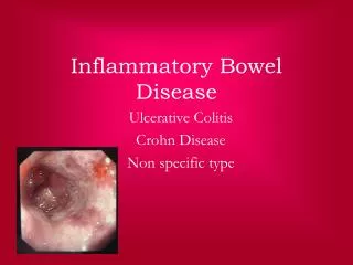
Inflammatory Bowel Disease
E N D
Presentation Transcript
Inflammatory Bowel Disease Case Scenario
Learning Objectives • Know the two forms of idiopathic inflammatory bowel disease (IBD). • Compare and contrast Crohn’sdisease and ulcerative colitis with respect to: a. clinical features and extraintestinal manifestations b. pathogenesis c. pathology (gross and microscopic features) d. complications (especially adenocarcinoma preceded by dysplasia)
A 56-year-old white male presented with a history of inflammatory bowel disease of 20 years duration and migratory polyarthritis. • Three months prior to admission, he experienced an exacerbation with a 40-pound weight loss and bloody diarrhea. • He was admitted to the hospital with complaints of 20 bloody diarrheal bowel movements per day, diffuse abdominal pain, and nausea and vomiting. • He stated that his flare-ups in the past were resolved with a short course of prednisone. He did not report any family history of inflammatory bowel disease.
On physical exam, the patient was a cachectic, no acute distress. His abdomen demonstrated positive bowel sounds and was nondistended, soft, and nontender. There was no hepatosplenomegaly or masses. • Rectal examination revealed black stool that tested positive for fecal blood, but no masses. The remainder of the physical exam was unremarkable.
On admission, the following lab results were noted: • His other laboratory values were unremarkable.
The patient was admitted to the hospital and treated with steroid therapy. There was no symptomatic improvement after eight days of therapy, so the patient was placed on NPO (nothing by mouth) and on TPN (total parenteral nutrition). He was transfused with two units of packed red blood cells. • Colonoscopicexamination revealed disease in the descending and sigmoid colon and rectum. Biopsies were performed; the interpretation by the pathologist was more consistent with ulcerative colitis than with Crohn disease. There was no evidence of dysplasia. • Options were discussed with the patient, and he decided to have a total colectomy
This large bowel has a diffusely inflamed mucosal surface, extending from the rectum to the cecum. Note the appendix for orientation. The right colon appears to be less involved; however, microscopically, the mucosa would show an active colitis. Is the picture consistent with Crohn disease? No, this is not consistent with Crohn’sdisease, because of the uninterrupted involvement of the mucosa. Although Crohn’sdisease may involve the colon, affected areas are sharply delimited by intervening unaffected areas.
The rectum shows a diffusely inflamed mucosal surface • Ulcerative colitis causes chronic diarrhea with grossly bloody, mucus-filled stools. Since the disease begins in the rectum and proceeds proximally without interruption (no skip areas), it is usually evident on proctoscopic exam, where pseudopolyps and ulcers can be seen.
What factors are believed to underlie the pathogenesis of inflammatory bowel disease (IBD)? • The etiology of inflammatory bowel disease (ulcerative colitis and Crohn disease) is unknown. • Familial aggregation suggests a genetic predisposition, but no definite genetic markers have been identified, except for the uniform presence of HLA-B27 in patients with IBD and ankylosingspondylitis. • Current opinion favors abnormal host immunoreactivity to luminal or mucosal antigens. • The beneficial effects of immunosuppressive therapy also point to the role of an aberrant immune response in the pathogenesis of IBD.
Colon, chronic ulcerative colitis, pseudopolyp • This image demonstrates a pseudopolyp (bulging mass of inflamed residual mucosa) surrounded by an extensive area of ulceration. Inflammatory infiltrate is seen in the submucosa.
What is the definition of a polyp? The term polyp is used to describe any nodule or mass that projects above the level of surrounding mucosa; it may be hyperplastic or neoplastic.
Why are the lesions in ulcerative colitis called pseudopolyps? These lesions are not really the result of mucosal proliferation, but rather are the result of mucosal ulceration. Focal areas that are unaffected by ulceration appear to project above the denuded mucosa surrounding them.
Colon, chronic ulcerative colitis • This image shows an ulcer base with fibrin, capillaries, and inflammatory cells. Note the distorted regenerating crypts at the edge of the ulcer. Compare the shape of the regenerating crypts with that of the normal crypts
How is the extent of these lesions different from that seen in Crohn disease?Crohn’sdisease causes transmural inflammation that ultimately results in fibrosis and thickening of the bowel wall.
Colon, chronic ulcerative colitis • The lamina propria shows an increase in acute and chronic inflammatory cells. A mucosal crypt is distended by a collection of neutrophils, known as a crypt abscess. Because ulcerative colitis is a diffuse mucosal disease process, crypt abscesses are common. Crypt abscesses are also seen in Crohn disease, but are not as common.
What histologic feature is seen in Crohn’sdisease that is not seen in ulcerative colitis? Granulomas and transmural inflammation in the resected specimen.
What are the complications of ulcerative colitis? • The most serious complication is the development of carcinoma. The cancers are preceded by dysplasia, which tends to arise in multiple sites. The risk of cancer is highest in patients with pancolitis of ten or more years duration, in whom it is 20 to 30 fold higher than in a control population. • Other life-threatening complications include: • severe diarrhea and electrolyte disturbances • severe colonic dilation (toxic megacolon) with potential for perforation and peritonitis • massive hemorrhage
Case scenario Presentation Ben has recurrent abdominal pain, weight loss and diarrhoea. He has been finding it difficult to manage at school because he finds his diarrhoea embarrassing. This has led to him missing school Medical history Ben is an 8-year-old boy who presented in primary care. There are concerns about his growth, his appetite is reduced and he is losing weight. He has been referred to secondary care for investigation On examination Ben is small for his age (height on the 9th centile) and quite thin (weight below the 2nd centile). He appears tired, is pale and is tender in the right iliac fossa
Next steps for management • Ben’s blood tests show that he is anaemic with a high platelet count, low albumin level and raised inflammatory markers (ESR and C-reactive protein) • Ben is referred for endoscopy, which shows patchy inflammation in the terminal ileum – a typical appearance of Crohn’s disease
Small and large intestines, Crohn’sdisease, regional enteritis - Gross, mucosal surface
Small and large intestines, Crohn disease, regional enteritis - Gross, mucosal surface • The specimen is a section of normal ileum, thickened ileum, and right colon. The intestinal wall is thick, the result of edema, inflammation, fibrosis, and hypertrophy of the muscularispropria. Linear ulcers are typically present in the diseased segment of bowel. • Unlike ulcerative colitis, the diseased areas are sharply demarcated from adjacent uninvolved bowel. • Note the area of normal colonic mucosa on the left and area of normal small bowel on the right. • In diseased bowel segments, the serosa is thickened and fibrotic, and often the mesenteric fat wraps around the bowel surface (creeping fat).
Are any other organs affected in Crohn’sdisease? In ulcerative colitis? Both Crohn disease and ulcerative colitis are systemic diseases, associated with varied extraintestinal manifestations of immunologic origin. These include polyarthritis, sacroiliitis, ankylosingspondylitis, uveitis, sclerosingcholangitis, erythemanodosum, and clubbing of the fingertips.
Bowel, Crohn disease • This low-power view shows focal areas of ulceration, submucosal edema, and transmural inflammation with lymphoid nodules. Fissures are narrow microscopic clefts that penetrate from the ulcer surface into muscle and are lined by granulation tissue. When the entire bowel wall is traversed, a sinus tract is formed that may open to the skin or adhere to an adjacent viscus
What is the risk of colon carcinoma in Crohn’sdisease? While the risk is increased, it is less so than in ulcerative colitis. There is a five- to six-fold increased risk over age-matched populations.
Bowel, Crohn’sdisease • At this magnification, crypt distortion, marked submucosal edema, fibrosis, and lymphoid aggregates can be seen. In addition, these patients may have noncaseatinggranulomas in the bowel wall. They are seen in about half of cases.
What are the complications of Crohn’sdisease? Fissures in the mucosa can extend through the wall and form sinus tracts, resulting in fistula formation to other loops of bowel, urinary bladder or vagina; there may be localized peritonitis and abdominal abscesses; fibrosis of the gut wall may lead to strictures and obstruction. Extensive involvement of the small bowel may cause marked loss of albumin (protein-losing enteropathy) or malabsorption.
In this view, marked thickening of the bowel wall can be appreciated. The thickening results from edema, inflammation, and fibrosis. In addition, a fissure penetrating the wall can be seen. This tracts of necrosis and acute inflammation start in the mucosa. Here, in cross section, the mucosal connection of the fissures cannot be seen. Frequently, fissures and sinuses resemble abscesses when they are cut transversely.
Bowel, Crohn disease, noncaseatinggranulomas • Noncaseatinggranulomas are present in the lamina propria of an uninvolved region of colonic mucosa. • Noncaseatinggranulomas are present in about half of Crohn disease cases in all tissue layers, both within areas of active disease and in uninvolved regions of the bowel. Granulomashave been documented throughout the alimentary tract, from mouth to rectum.
The worst case scenario • The first indication that something was off in my life was when I got a bartholin gland cyst. I years later attribute this to crohns. But why is my crohns attacking my vaginal area. The fistulas... At first they were on the cheek now they are getting closer and closer to my vaginal opening they meaning about 4 holes. I hate examining myself. I think it makes me more depressed to see them