Download
1 / 82
850 likes | 1.41k Vues
Acute Abdominal P ain in the Tropics. 2011 Global Health Missions Conference Louisville, Kentucky Presented by Bruce C. Steffes, MD, FACS, FWACS, FCS(ECSA) Certificate of Knowledge in Clinical Tropical Medicine and Travelers Health (ASTMH). Introduction.

E N D
Acute Abdominal Pain in the Tropics • 2011 Global Health Missions Conference • Louisville, Kentucky • Presented by • Bruce C. Steffes, MD, FACS, FWACS, FCS(ECSA) • Certificate of Knowledge in Clinical Tropical Medicine and Travelers Health (ASTMH)
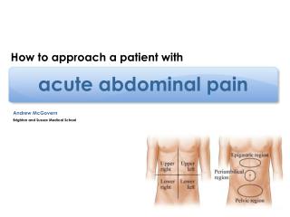
Introduction • Surgery in the developing world is different • Different diseases or at least different prevalence • Advanced pathology • Fewer care givers • Limited resources
Diseases seen less commonly in the developing world • Diveriticulitis • Acute and chronic cholecystitis • Appendicitis • Small bowel obstruction due to adhesions
Diseases seen more commonly in the developing world • Primary peritonitis • Perforated duodenal ulcers • Volvulus • Adult intussusception • Tuberculous peritonitis • Pigbel
What are diagnoses seen in the two-thirds world? • University Hospital in Ghana1 • Appendicitis • Perforated typhoid • Bongolo Mission Hospital, Gabon2 • Incarcerated/strangulated hernias • Appendicitis • Volvulus • Adhesive SBO • Perforated typhoid • Tenwek Mission Hospital3 • Volvulus • Appendicitis • Perforated PUD • Trauma • Perforated typhoid • SBO
Case Presentation • 5 yo boy from Papua New Guinea. • Pig feast 5 days before • Severe abdominal pain 4 days during with fever, nausea & diarrhea. Intermittent cramps, especially with eating & drinking • WBC 14,400 • Abdomen initially soft. • Dx? Tx?
NPO, nasogastric tube and antibiotics • “Dark” NG output, “dark” diarrhea and abdomen became “surgical” Dx?
Pigbel • (Enteritis Necroticans, Necrotizing Enteritis) was reported first in medieval Europe and again in Germany after WWII when it was called “darmbrand” (gut-fire). It resurfaced in the early 1960s in Goroka, PNG with culture-positive cases and was the most common cause of death in children >24 months). At one time, Pigbel was the most common cause of acute abdominal pain in the PNG Highlands(Prior to vaccination was the most common cause for abdominal laparotomy in that area)
Incidence of Pigbel • Male > Female (2.2:1) – probably because males encouraged to eat more protein for strength • 70% between ages 1 – 10. Infants protected by maternal antibodies. Can be seen in young adults (25%) • More common in dry season (better weather leads to more frequent pig feasts)
Bacteriology of Pigbel • Clostridium Perfringens Type C (also known as Clostridium welchii) = Anaerobic, gram +, spore-forming rod - found in human stool, pig stool and soil • Spores are heat stable up to 95 C (Boiling point of water is 95 in the PNG Highlands) • Type A commonly causes food poisoning (botulism). Type C grows in protein-rich chyme and produces a b – toxin).
Effect of the b-toxin • b-toxin is rapidly degraded by intestinal proteases (e.g. trypsin) in well-nourished people • The toxin attacks the intestinal lining and causes inflammation and necrosis and may also cause arterial thrombosis
How It Comes Together • Trypsinhas low activity • Malnutrition causes a decrease in the pancreatic production of all proteases including trypsin, a key enzyme in the digestion of meat and protein and the toxin • Ascaris infestation and diet rich in sweet-potato (kaukau) cause high levels of heat-stable trypsin inhibitors • Contamination risk is high • Unwashed hands and feet of food handlers • Poorly cooked pork/meat or spillage of pig’s bowel contents in mumu preparation • Sporadic high protein meals provides growth medium for C.P.
Pathology • Blood and pus in stool (from “sloughing” enteritis of jejunum, ileum and colon) • Transmural infection of the bowel (patchy segmental ulcerative necrosis) • Gas gangrene, separation of the layers of the bowel wall, pseudomembranes • Affects jejunum > ileum > cecum > colon
Types of Pigbel: • Mild Diarrheal (type IV) • May go undiagnosed or diagnosed as gastroenteritis (GE) • Usually only diarrhea but can progress to Type IV • Mortality: Rare • Subacute Surgical (type III) • Presents later • Complication of Type II (See next category) • Mortality: 49%
Types of Pigbell • Acute Surgical (type II) • Present with ileus, small bowel obstruction (SBO), strangulation, perforation, peritonitis • Mortality: 42% • Acute Toxic (type I) • Fulminant toxemia and shock • Usually young children (immunologically naïve) • Mortality: 85% (Some deaths before hospital)
The Clinical Course • Symptoms usually become apparent 48 hours after a large meat or protein meal but can present up to a week later • Present with colicky or constant abdominal pain, vomiting with dark emesis (blood flecks), blood in stool, foul flatus, and diarrhea early • Tachycardic, febrile, dehydrated, tender & distended upper abdomen with visible bowel, guarding, rigidity, decreased bowel sounds
The Clinical Course • Symptoms consistent with ischemia • Pain out of proportion • Eating may increase pain • Late symptoms: partial SBO, malnutrition, fibrosis, adhesions, malabsorption and strictures (especially with Type III) • Mortality due to peritonitis, septicemia, dehydration, electrolyte abnormalities, and shock
Diagnostic Approach • High index of suspicion • Early recognition of pigbel and quick action are of utmost importance. The toxin begins attacking the bowel instantly and constantly. Timely recognition and treatment may reduce severity or even prevent death of the child! Early fluid resuscitation, decompression of SB, and appropriate antibiotics may preclude need for laparotomy. If severe, early referral and surgery may prevent death.
Diagnosis of Pigbel • Gas in bowel wall or SBO on abdominal x-ray • Bloody NG aspirate or blood in stool • Neutrophilic leukocytosis (>/= 20,000) • Serological test possible, ? availability (immuno-florescence using type C coated silicon beads) • Culture C.P. from stool (anaerobic blood agar) • Bloody ascites on ultrasound
Treatment of Pigbel • Correction of fluid and electrolyte deficits; hydrate well; correct moderate to severe anemias. • Nasogastric drainage • Intravenous antibiotics - CMP, Crystalline PCN and Metronidazole/Tinidazole (+/- Gentamicin)
Treatment of Pigbel • Treatment of Ascaris • ?treatment of malaria • Consider hyperalimentation or TPN if course prolonged • Antiserum +/- (Not readily available or effective)
Early Hospital course • If improves, wait 24 hours then slowly go from oral rehydration solution (ORS) milk solids • After 48 hours , laparotomy if there if failure to improve: high NG output, persistent SBO by x-ray, persistent peritonitis, high white count, persistent fever • Acute decompensation requires emergent surgery
Surgery • Due to the rapid progression of pigbel, the decision for surgery is often a judgment call by the surgeon based on clinical experience • Urgent laparotomy with wide resection of SB to normal margins • Questions that arise: • How much bowel to resect? • End-to-end anastomosis vs. 2ostomies • Which patients to do second look?
Surgery Findings • Palpable loops of thick bowel • Enlarged mesenteric nodes typical • “Tiger Striping” • “Skip Lesions” • Mucosal Ulcerations • Perforations/SBO
Post Op Care • Strict I & O, Adequate fluid resuscitation and good nursing care. • Attention to the CBC (transfusion is needed) and K+ levels • Nutritional supplement
Prevention of Pigbel • Type C toxoid immunization. Inactivated toxin: 0.5 cc given at 2, 4 and 6 months of age with the DPT vaccine. Protects 2-4 years • Was used from 1980- mid 1990’s and cases were 1/5 of pre-immunization levels, When the PNG government felt it was too expensive (and quit paying for it), it became an orphan drug and the manufacturer quit making it. • In one recent study, 6 of 25 non-immunized kids had pre-existing antibodies to C.P. type C indicating that the organism is still common.
Prevention of Pigbel • Changes in dietary habits (Less reliance on sweet potatoes and more regular protein) • Changes in cooking methods (higher temperatures) and better preservation of food • Changes in hygiene and food preparation • Public Health Education • Eradication of Ascaris
Case Study • 10 yo Togolese boy presents in the dry season with history of fevers and malaise. He has had intermittent nausea and mild diarrhea. He was treated for malaria. He worsened 48 hours ago and hasn’t eaten since then. • Dx, Rx?
Typhoid fever • Cause: • Salmonella entericaserovartyphi • Certain non-typhoid salmonella (NTS), particularly Paratyphoid strains A, B, C. • Disease of poor sanitation, often seasonal
Typhoid Fever Bitar & Tarpley, Reviews of Infectious Diseases 7:257-271, 1985
Clinical Features of Typhoid • Classically a four week disease • Weeks one and two: fever, headache, abdominal pain • Week three: “typhoidal state” with disordered mentation and toxemia • Week four: Defervescence and improvement
Lab in Typhoid Disease: • Leukopenia/thrombocytopenia are common • Culture is the best diagnostic tool – but may not be available.
Widal? • Widal test: very controversial • Conclusion of a paper by Tupasi et al ([Phil J Microbiol Infect Dis 1991, 20(1):23-26] “Culture isolation of Salmonella typhi from blood and bone marrow should be considered the standard diagnostic test to confirm typhoid fever. A single Widal test in an endemic area is of no diagnostic value. In addition, it should not be used as a screening test in asymptomatic individuals. Neither should a "negative" Widal test rule out the diagnosis of typhoid fever in patients with signs and symptoms of the disease since a "negative" Widal test may be seen early in the course of illness. The Widal test should not also be used as the basis for deciding the duration of antimicrobial therapy.”
If Surgery Indicated Emergently • Aggressive resuscitation prior to OR with appropriate antibiotic coverage (triple antibiotics to cover GI flora as well as Salmonella) and sharing of the Gospel • Ampicillin and chloramphenicol are no longer the drugs of choice. Fluoroquinolones (?decreasing efficacy) and third generation cephalosporins are probably the best at present.
Indications for Surgery in Enteric Fever • Surgery for carrier state is NOT a usual indication. Normally, do only for chronic cholecystitis per se (doesn’t always work for carrier state) • Hemorrhage (1.5 - 10% of patients, bleeding usually in 3 or 4th week, usually UGI in type and may be hard to find if in the small intestine) • Perforation (1 - 5% of patients, common in the second and third weeks of illness, but can be much later. Some patients perforate without an obvious prodrome) • Mortality for perforation is as high as 40%, affected by many factors in the austere environment.
Indications for surgery: • Pneumoperitoneum on x-ray (may require left lateral film) • Persistent palpable mass (especially with erythema of abdominal wall) • Diffuse peritonitis or positive peritoneal tap • Persistent sepsis/failure to improve on medical therapy • Suspicious of abdominal catastrophe but negative x-rays? Do frequent examinations (by the same or equally experienced examiner) and x-rays (q. 6 h at first) until improvement or perforation is evident.
Multiple perforations Up to 3 or so – oversew Multiple – consider resection with single anastomosis Aggressive peritoneal debridement and/or irrigation of the peritoneal cavity Surgical Approach
Consider retention sutures Consider a second-look operation A negative laparotomy is rare and better tolerated than a missed perforation. Surgical Approach
Typhoid Cholecystitis • Acute cholecystitis – very uncommon • Predominance in children • Often advanced (gangrene or perforation) because of low index of suspicion Courtesy, Dr. J. Brown, Cameroon
Typhoid Cholecystitis • Acute cholecystitis – very uncommon • Predominance in children? • Often advanced (gangrene or perforation) because of low index of suspicion
Bowel Obstruction • The differential diagnoses have different probability • Examples: • Decreased adhesions due to fewer surgeries (except in women where PID increases risk of adhesions) • Lower frequency of colon cancer and diverticulitis • Increased prevalence of lymphogranulomavenereum stricture
Case • 8 year old female presents with abdominal swelling, pain and vomiting. WBC 14,200. 6% eosinophilia. Hg 8.9 gm%. A sausage-shaped RLQ mass was palpated.