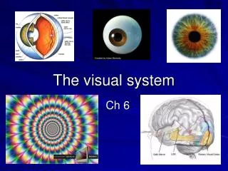
Topic 11 The Central Visual System Lange
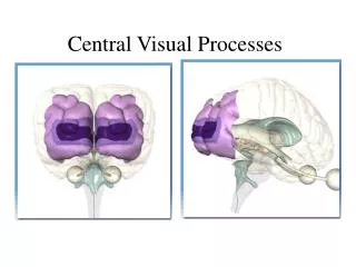
Topic 11 The Central Visual System Lange. Biology 463 - Neurobiology. The Retinofugal Projection. The retinofugal projection consists of the Optic Nerve, Optic Chiasm, and Optic Tract. The Retinofugal Projection. Right and Left Visual Hemifields

Topic 11 The Central Visual System Lange
E N D
Presentation Transcript
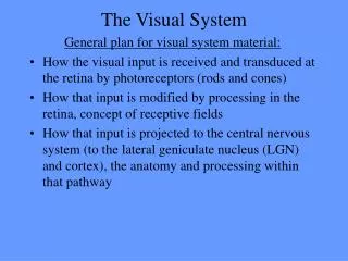
Topic 11 The Central Visual System Lange Biology 463 - Neurobiology
The Retinofugal Projection The retinofugal projection consists of the Optic Nerve, Optic Chiasm, and Optic Tract
The Retinofugal Projection • Right and Left Visual Hemifields • Left hemifield projects to right side of brain and right to the left
Visual deficits from lesions in the retinofugal projection are shown in these images. Notice the specific types of loss shown in black as compared to the intact fields
Nonthalamic Targets of the Optic Tract are areas that will use visual input but not in the sense of SEEING OBJECTS. • Hypothalamus: Biological rhythms, including sleep and wakefulness • Pretectum: Size of the pupil; certain types of eye movement • Superior colliculus: Orients the eyes in response to new stimuli X X X
The Lateral Geniculate Nucleus (LGN)The pathways occur in alternating layers.Contralateral = on the other sideIpsilateral = on the same side
Parvocellular Cells • Parvocellular cells, also called P-cells, are neurons located within the parvocellular layers of the lateral geniculate nucleus (LGN) of the thalamus. • "Parvus" means "small" in Latin, and the name "parvocellular" refers to the small size of the cell compared to the larger magnocellular cells. • The parvocellular neurons are sensitive to color, and are more capable of discriminating fine details than their magnocellular counterparts. Parvocellular cells have greater spatial resolution, but lower temporal resolution, than the magnocellular cells. Magnocellular Cells • Magnocellular neurosecretory cells are large cells within the supraoptic nucleus and paraventricular nucleus of the hypothalamus. • There are two types of magnocellular neurosecretory cells, oxytocin- producing cells and vasopressin-producing cells, but a small number can produce both hormones. • These cells are neuroendocrine neurons, they are electrically excitable, and generate action potentials in response to afferent stimulation.
The Lateral Geniculate Nucleus (LGN) Inputs Segregated by Eye and Ganglion Cell Type P type = (also known as beta or midget ganglion cells) are believed to be responsible for detecting details in vision. M type = (also known as alpha or parasol ganglion cells) are believed to be responsible for detecting motion. nonM-nonP type =are a diverse group of cell types that make up the remaining 5% of RGCs. Their roles in vision are less understood than M- and P-type ganglion cells, but it is known that some non-M, non-P type cells are involved in color vision.
Anatomy of the Striate Cortex The calcarine sulcus is where the primary visual cortex is concentrated. The central visual field is located in posterior portion of the calcarine sulcus and the peripheral visual field in the anterior portion.
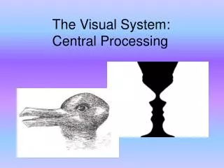
Anatomy of the Striate Cortex Retinotopy • Map of the visual field onto a target structure (retina, LGN, superior colliculus, striate cortex) • Central visual field overrepresented • Discrete point of light: Activates many cells in the target structure due to overlapping receptive fields • Perception: Based on the brain’s interpretation of distributed patterns of activity
Anatomy of the Striate Cortex Inputs to the Striate Cortex • First binocular neurons found in striate cortex - most layer III neurons are binocular (but not layer IV)
Anatomy of the Striate Cortex Outputs of the Striate Cortex: • Layers II, III, and IVB: Projects to other cortical areas • Layer V: Projects to the superior colliculus and pons • Layer VI: Projects back to the LGN
Anatomy of the Striate Cortex Cytochrome Oxidase Blobs • Cytochrome oxidase: mitochondrial enzyme used for cell metabolism • Blobs: Cytochrome oxidase staining in striate cortex • Each blob centered on an ocular dominance column in layer IV • Receive koniocellular inputs from LGN
Physiology of the Striate Cortex • Monocular Receptive Fields • Layer IVC: Similar to LGN cells • Layer IVC: Insensitive to the wavelength • Layer IVC: Center-surround color opponency • Binocular Receptive Fields • Layers superficial to IVC: First binocular receptive fields in the visual pathway • Two receptive fields - one for each eye
Physiology of the Striate Cortex Cortical Receptive Fields • Orientation Selectivity
Physiology of the Striate Cortex Cortical Receptive Fields • Direction Selectivity • Neuron fires action potentials in response to moving bar of light
Physiology of the Striate Cortex Cortical Receptive Fields • Simple cells: Binocular; Orientation-selective; Elongated on-off region with antagonistic flanks responds to optimally oriented bar of light • Possibly composed of three LGN cell axons with center-surround receptive fields
Physiology of the Striate Cortex Cortical Receptive Fields • Complex cells: Binocular; Orientation-selective; ON and OFF responses to the bar of light but unlike simple cells, no distinct on-off regions
Physiology of the Striate Cortex Cortical Receptive Fields • Blob Receptive Fields: • Circular • Monocular • No orientation or direction selectivity • Majority of color-sensitive neurons outside layer IVC • Specialized for analysis of object color
Physiology of the Striate Cortex Parallel Pathways: Magnocellular; Koniocellular; Parvocellular
Physiology of the Striate Cortex Cortical Module • Each module capable of analyzing every aspect of a portion of the visual field