The visual system
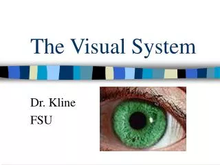
The visual system. Macleod’s 14 th edition – chapter 8 Adonia haddad. Eye ..in Bony orbit Protection .. Eyelid ( from injury / maintain tear film)


The visual system
E N D
Presentation Transcript
The visual system Macleod’s 14th edition – chapter 8 Adonia haddad
Eye ..in Bony orbit • Protection .. Eyelid ( from injury / maintain tear film) -Upper lid elevated : by levatorpalpebraesuperiosis.. Cranial Nerve lll& Mular muscle.. nerve sympathetic autonomic system - eyelids Closure : orbicularis oculi .. cranial neve vii • Orbits contains six extraoclear muscles : 1- superior rectus 2-medial rectus 3-lateral rectus 4-inferior rectus 5- superior oblique 6-inferior oblique • Orbit houses : 1- lacrimal gland 2-blood vessels 3-autonomix nerve fibers 4-cranial nerves (ll , lll , lV , Vl) • The contents are cushioned by orbital FAT ( enclosed anteriorly by the orbital septum and eyelids ) • Conjictiva : thin mucous membrane lining the posterior aspects of the eyelid ,, reflected at the superior and inferior fornices on the surface of the globe . Coated in a tear film that protects and nourishes the oclear surface Anatomy and physiology
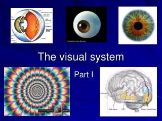
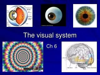
25mm ( length) • 3 distinct layers : 1- outer fibrous layer :sclera and cornea ( cornea 2/3 of reflective power of the eye .. Focusing incident light on the retina ) 2- middle vascular layer ( uveal tract) : ciliary body and the iris ( anteriorly) .. the choroid (Posteriorly) 3- inner neurosensory layer : retina (converts light to neurological signals ) Eye :
6 extraoclear muscles • Responsible of eye movement • Cranial nerve lll … superior/medial/ inferior rectus muscle and inferior oblique muscle • -Cranial nerve lV .. Superior oblique mucle • - cranial nerve Vl .. Lateral rectus muscle Cranial nerves originate in the misbrain and pons and then pass through the cavernous sinus EXTRAOCLEAR MUSCLES :
Tear film/ cornea/crystalline lens .. the major refractory elements • cornea .. The greatest refractive power and the main refracting element of the eye • lens .. Additional controllable refraction , causing the light to focus on the retina • When light is : • Precisely focused on the retina … emmetropia • Focus point falls behind the retina .. Hypermetropia ( long sightness) .. Eye is too short • Focus infront of the retina .. Myopia (short sightedness) .. Eye is too long These errors (2 and 3 ) can be corrected with 1)Lenses .. For point num 2 use a convex (plus) lens & for point num 3 use a concave (minus ) lense 2)pinhole Refractive elements of the eye :
Consists of : • Retina / optic nerve / optic chiasm / optic tracts / lateral geniculate bodies/optic radiations / visual cortex • Any defict in this pathway leads to specific field defects Visual pathway :
Pupil … Control the amount of light entering the eye • Intensity of the light determines the aperture of the pupil .. Via autonomic reflexes : 1)Pupillary constriction : by parasympathetic nerves .. The afferent pathway is the optic nerve - synapsing in the pretectal nucleus of midbrain --- axons synapse in both cranial nerve lll (edinger – westphal ) nuclei before passing along the inferior devision of the oculomotor nerve --- to synapse in the ciliary ganglion .. The efferent postganglionic fibers pass to the pupil vial short ciliary nerves --- result in constriction 2) Pupillary dilation : by sympathetic nerves .. Originates in the hypothalamus - passing down to the ciliospinalcentre of budge at level of T1 --- fibers then pass to and synapse in the superior cervical ganglion before joining the surface of the internal carotid artery ---- and pass to the pupil along the nasociliary and the long ciliary nerves Pupillary pathway
Bear in mind the anatomy of te eye and the visual pathway • This will enable you to work from front to back to include and exclude differential diagnosis • COMMON PRESINTING SYMPTOMS : Start with open questions allow the patient to describe the condition in their own words Provide clues for more directed questions later 1)ALTERED VISION Vision altered by intraoclear diseases which leads to : • change in the optical or refractive properties of the eye • Prevent the incident light rays from being clearly focused on the retina Alternatively, it may results from extraoclear factors associated with : 1)damage to the visual pathway , which runs from the optic nerve to the optical lobe History:
Establish whether the change in vision is : • Sudden 2) gradual .. Both of them have their own specific set of differential diagnosis • vision may be not just reduced but also distorted … this results from disruption to the normal structure of the macula , the central part of the retina .. The most common cause is macular degeneration but also may frequently stem from an epiretinal membrane ,vitreous traction or central serous retinopathy • Flashes and floaters … This results from disturbance of the vitreous and the retina , occurring most commonly in posterior vitreous detachment Ususally found in older patient as the vitreous gradually degenerates and liquefies , causing it to peels from the retina The vitreous is attached to retina in certain regions , in these regions the vitreous either : 1)detaches with traction … resulting in flashing light 2) Detaches by tearing the retina .. Releasing retinal pigment cells Patient will see either of these as floaters
Haloes : are colored lights seen around bright light Occurs with corneal edema and mostly associated with angle-closure glaucoma What to ask about when you have a patient with altered vision : 1)Did it start sudden , or gradually ? 2)How the vision is affected (loss/cloudy/floaters / distortion)? 3) One or both eyes that are affected ? 4) The whole or only part of the visual field affected ? 5) If partial . Which part of the visual field is affected ? 2)Pain Ask: 1)when it began ? 2)Anything started the pain 3)Character of the pain 4)severity? 5)Exacerbation and relieving factors 6)Associated symptoms
Cornea is one of the highly innervated regions of the body When corneal nerves are activated … leads to 1)Pain 2)sensation of a foreign body 3)reflex watering 4) photophobia And there are causes will be mentioned later 3)Red eye The eye is covered with a network of vessels in the conjunctiva ,episclera and sclera Ciliary vessels are also found around cornea • causes leads to red eye : 1)Dilation or hemorrhage in these vessels 2)uveitis , in acute angle closure glaucoma and corneal irritation … the ciliary vessels around the cornea become more prominent ( ciliary flush) The appearance is distinct from conjunctivitis , in which there is blanching of vessels towards the cornea
Ask: 1)If the eye is painful or photophobic? 2)Vision is affected? 3)Recent trauma? 4)Eye is itchy? 5)Any discharge? 6)Any recent contact lens wear or foreign body exposure? There are causes will be mentioned later 4)Double vision (diplopia) Whether it is : 1)Monocular .. Results from intraocular disease in one eye 2)Binocular .. Results from imbalance in eye movement There are of causes will be mentioned later Ask: 1)Occurs ijn one or both eyes? 2)Character ? Whether Images seen side by side ? One above the other? Or at angle ? 3)Associated with trauma
Test the eye movement and use your knowledge of the functions to of the extraoclear muscles to work out whch cranial nerve is affected in binocular diplopia • 5)discharge Increase in discharge from the eye results from 1)Increase in production 2) Decrease in drainage from the oclear surface • Irritation of cornral nerves activates cranial nerve Vl and results in reflex tearing response Tears normally drain through the punctum at the medial end of the lower eyelid into nasallacrimal duct .. Which opens below the inferior turbinate into the nasal cavity Blockage of tear drainage or abnormal lid position can also result in excessive drainage Ask: 1)Discharge clear?opaque? 2)Associated pain ?foreign body sensation? Itchiness? 3)Any noticed abnormalities ? Such as red eye ? There are causes will be mentioned later
6)swollen eyes Orbit is an enclosed structure except anteriorly Any swelling will results in : 1)Anterior displacement of the globe 2) Proptosis Ask • unilateral or bilateral • Acute ? Chronic ? • Painful? • Itchiness? Irritation ? • Associated with double vision? There are causes will be mentioned later
Past ocular history Ask 1)any known opthalmic conditions 2)Having amblyopia (reduction in vision in one eye from childhood) .. this may limit best-corrected visual acuity 3) if the patient wears glasses or contact lenses 4)Last time they had their eyes checked for refractive correction 5)Any previous eye operation that may affect vision • Past medical history : • Focus on systematic diseases that could affect the eyes ( directly / side effect of treatment ) : 1)diabetis or HTN .. Especially in the context of visual loss of double vision 2)asthma or COPD orperipheral vascular disease if starting glaucoma medications • Drug and allergy history The eyes may be affected by medications for other conditions .. Like glaucoma exacerbation by conjunctival absorption of nebulished anticholinergic drugs in copd Medications for the eyes ( like beta blocker eye drops )can aggrevate other conditions like asthma Ask: 1)History of fever or allergy .. If the patient has itchy eyes
Family history Several eye diseases have an inherted predisposition Ask: 1)History of glaucoma in first order relatives 2)Genetic diseases affecting the eye ... Includes retinitis pigmentosa 3)Thyroid eye disease … may have positive family history of autoimmune disease Social histoty : Visual impairment has a wide range of effects on daily life Ask: 1)Dailly activity require good vision … reading/Tv /sport/hobbies/… 2)Driving 3)Occupation .. Certain professions including drivers of heavy good vehicles / pilots requirespecific visual acuity criteria 4)Smokingband alcohol use… this may affect vascular and optic nerve function within the eyes
General examination : Examine carefully and systematically 1)Posture and gait 2)Head position 3)Facial asymmetry and dysmorphic features 4)Eyelid position and periocular skin 5)Position and asymmetry of gaze (any squint/strabismus) • Visual aquity: Mandetory to assess Each eye should be assessed separately .. The most common method of testing distance visual acuity is using snellen chart ( displays a random selection of letters at diminishing font size in successive lines ) Ask: 1)To wear their distance spectacles if thtey require them .. Near/ reading spectacles should be worn only when testing reading vesion Physical examination
Pupils : Examine 1) inspect generally for squint and ptosis 2)Examine pupil shape and symmetry .. Physiological anisocoria (unequal pupil size ) is seen in 20% of population .. Eyes should be assessed te determine which is the abnormal pupil
Relative afferent pupillary defect (RAPD) • is an important clinical sign that occurs when disease of the retina or optic nerve reduces the response of the eye to a light stimulus • Testing for RAPD is an extention of the direct and consensual light responses