The Human Visual System
The Human Visual System. The Eye. In this section . . . . Anatomy of human eye Image formation by human eye Method of light detection Retinal processing Eye optical defects and diseases. Human Visual System. Detection. Processing. Exposure Control. Image formation. Retina Rods

The Human Visual System
E N D
Presentation Transcript
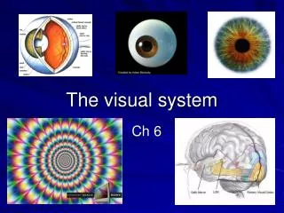
The Human Visual System The Eye
In this section . . . • Anatomy of human eye • Image formation by human eye • Method of light detection • Retinal processing • Eye optical defects and diseases
Human Visual System Detection Processing Exposure Control Image formation • Retina • Rods • Cones • Brain • Cornea • Lens • Iris/pupil • Photoreceptor • sensitivity
Human Eye Ciliary Muscle Sclera Ear side (Temporal) Iris Vitreous Humor Fovea Pupil Eyelens Retina • Human eye is a complete imaging system. Optic Nerve Cornea Nose side (Nasal) Aqueous Humor Choroid Suspensory ligament
Image Formation Object Image • The curved surfaces of the eye focus the image onto the back surface of the eye.
Cornea Sclera Cornea • The outer wall of the eye is formed by the hard, white sclera. • Cornea is the clear portion of the sclera. • 2/3 of the refraction takes place at the cornea.
Iris and Pupil Iris • Colored iris controls the size of the opening (pupil) where the light enters. • Pupil determines the amount of light, like the aperture of a camera. Pupil Iris open Dilated pupil Iris closed Constricted pupil
Lens Ciliary muscle • Eye lens is made of transparent fibers in a clear membrane. • Suspended by suspensory ligament. • Used as a fine focusing mechanism by the eye; provides 1/3 of eye’s total refracting power. • Non-uniform index of refraction. Lens Suspensory Ligament Transparent Fibers Cross section of the eye lens
Accommodation Distant object • The suspensory ligaments attach the lens to the ciliary muscle. • When the muscle contracts, the lens bulges out in the back, decreasing its focal length. • The process by which the lens changes shape to focus is called accommodation. Relaxed muscle Taut ligaments Near object Contracted muscle Slack ligaments
Aqueous Humor and Vitreous Humor • Transparent gelatinous liquid filling the eye. • Provides nutrients to the cornea and eye lens. • Also helps maintain the eyeball shape with its pressure. Vitreous Humor Aqueous Humor
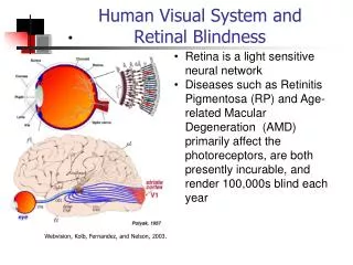
Retina • Retina is the photosensitive “detector” for the eye. • Two types of receptors in the retina: rods for low light level, and cones for color. • Located at the center of the retina, fovea contains a greater concentration of cones. • Signals from the receptors leave through the optic nerve tothe brain. Retina Fovea Optic Nerve
Plexiform Layer • The retina is made of three layers: • Plexiform layer is a network of nerves which carry the signals from the photo receptors. • Photo receptors. • Choroid provides nourishment to the receptors, as well as absorb any light that didn’t get absorbed by the photo receptors, like a antihalation backing in film. Fovea Photo receptors Light Plexiform Layer Choroid Optic Nerve
Rods and Cones Synaptic endings • Highly sensitive to low light level or scotopic conditions. • Black and white. • Dispersed in the periphery of the retina. Cell nucleus Inner segments Outer segments Rod Cone • Sensitive to high light level or photopic conditions. • Three types of cones responsible for color vision. • Concentrated in the fovea.
Adaptation • Why can’t you see immediately after you enter a movie theater from daylight? • The threshold of detection changes with overall light level. • The switch is quite gradual, until the sensitivities of cones and rods cross over at about 7 minutes in the dark. Photopic (cones) Scotopic (rods) Threshold of detection (log scale) 0 5 10 15 20 25 30 Time in dark (minutes)
80º 80 º 60 º 60 º 40 º 40 º 20 º 20 º 0 º Distribution of Photoreceptors Visual Axis • Cones are concentrated in the fovea. • Rods predominate the periphery. • There is a blind spot where there are no photoreceptors, at the point where the nerves exit the eye (optic nerve). Temporal Nasal Blind spot 160 140 Rods 120 Number of receptors per mm2 100 80 60 40 Cones 20 40 º 60 º 20 º 40 º 20 º 0 º 60 º 80 º Angle
S I L Relative response 400 460 490 500 530 600 650 700 Wavelength (nm) Blue Cyan Green Red Human Vision • Human Cone Response to Color • three cone types (S,I,L) correspond to B,G,R
Retina • The retina is made of network of nerve cells. • The network works together to reduce the amount of information in a process called lateral inhibition. Cones Light Rods Bipolar cells To optic nerve Amicrine cells Ganglion cells Horizontal cells
Hermann Grid • Illustrates lateral inhibition.
A B Hermann Grid • Point A looks darker because there are 4 inhibitory inputs • Point B looks lighter because there are only 2 inhibitory inputs
Mach Bands Actual brightness Perceived by you
Eye Defects Object at infinity • Image focuses on the retina for a normal eye. • Distant objects look blurry for a myopic (near sighted) eye. • Near objects look blurry for a hyperopic (far sighted) eye. Normal Myopic Hyperopic Eyes at relax state.
Myopia - Near sightedness • Distant objects look blurry because the eye cannot relax any farther so that the image is focused before the retina. • Near object in focus without accommodation. • Corrected with a negative lens. Far object Myopic eye relaxed Blurry Near object Myopic eye relaxed In focus Far object Myopia corrected with a negative lens The virtual image from the diverging lens appears to be closer.
Hyperopia - Far sightedness Far object • Near objects look blurry because the eye cannot accommodate enough for near objects. • Far object in focus. • Corrected with a positive lens. Hyperopic eye Partially accommodated In focus Near object Hyperopic eye Fully accommodated Blurry Near object Hyperopia corrected with a positive lens Light from the converging lens looks as though it is coming from the distance.
Contact Lens • Contact lens is an alternative to corrective lenses. • Changes the curvature of the cornea by adhering to the surface with some fluid. Contact lens Cornea Fluid
Presbyopia - “Old eye” • Lens hardens with age. • Eye cannot adequately accommodate near objects. • Bifocals (lens with two focal lengths) contains a concave lens for distance (if needed) and a convex lens for near objects. Concave for distance correction (if needed) Convex for near object correction Far objects Near objects magnified
Astigmatism • The cornea is not spherical; Focal length different from one plane to a perpendicular plane. Cornea Object F’ horizontal F’ Vertical Direction of blur Image at F’ Horizontal Image at F’ Vertical
Astigmatism • Correction of astigmatism is done through the use of a cylindrical lens. • Cylindrical lens converge rays in one plane but not the perpendicular plane. Cylindrical lens Rays in the vertical plane are undeviated Rays in the horizontal plane are focused
Common Eye Diseases • Cataract - Clouding of the lens. • Symptom: Loss of vision • Cure: Lens replacement • Glaucoma - Pressure buildup in the eye, damaging the retina. • Symptom: Loss of vision first in the periphery. • Cure: Surgery to drain fluid from the eye. • Loss of vision is usually permanent
Common Eye Diseases • Detached retina - portion of the retina detaches from the back of the eye. • Symptom: Perception of flashes, Loss of vision • Cure: Laser surgery to reattach retina • Pink eye - Infection of the surface of the eye. • Symptom: Irritation • Cure: Antibiotics Ow!
Your eye care Go see a doctor if you think there is something wrong with your eye- Early detection is essential to keeping damage low and preventing permanent loss of your vision.