Autoimmune Diseases
Autoimmune Diseases. Dr. Raid Jastania. Autoimmune Diseases. Group of diseases with common pathological process Presence of auto-antibody ?defect in B-cells or T-cells Genetic and Environmental etiological factors Result in failure of self-tolerance

Autoimmune Diseases
E N D
Presentation Transcript
Autoimmune Diseases Dr. Raid Jastania
Autoimmune Diseases • Group of diseases with common pathological process • Presence of auto-antibody • ?defect in B-cells or T-cells • Genetic and Environmental etiological factors • Result in failure of self-tolerance • The process involve Ag-Ab binding and Ag-Ab complex formation • Process may lead in complement activation and inflammation with tissue injury
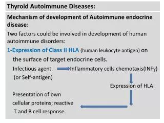
Autoantibody • Failure to maintain self-tolerance • Autoantibody can be formed to: • Nuclear antigen • Cytoplasmic antigen • Cell surface antigen • Proteins and phospholipids
Anti Nuclear Antibody (ANA) • ANA represent diversity of antibodies that bind to several nuclear antigen • Anti-DNA • Anti-Histone • Antibody to Non-Histone • Antibody to nucleolar antigen • ANA is usually detected by indirect immunofluorescence
Anti Nuclear Antibody (ANA) • Patterns of ANA staining: • Homogenous: antibody to chromatin, histone or DNA • Rim/Peripheral: antibody to DNA • Speckled: antibody to histone (Sm, ribonucleoprotein RNP, SS-A (Ro), SS-B (La)) • Nucleolar pattern
Systemic Lupus Erythematosus • Autoimmune Disease • Multi-system disease • Variable behaviour, unpredictable, remitting relapsing, acute and gradual. • May involve any organ • Common: Skin, kidney, serosal membranes, joints, heart.
Systemic Lupus Erythematosus • ANA (anti-DNA, anti-Sm, anti-phospholipid) • Prevalence: 1/2500 person • Female: male 9:1 • 1/700 women • More common and severe in blacks (1/245) • Onset: 2nd or 3rd decade
Malar Rash Discoid Rash Photosensitivity Oral ulcers Arthritis Serositis Renal disorder Neurologic disorder Hematologic disorder Immunologic disorder ANA SLE: Criteria for Diagnosis
SLE • ANA is sensitive to SLE but not specific • ANA is present in 5-15% of normal people • More specific to SLE are” • Anti-DNA • Anti-Sm • LE bodies (hematoxylin bodies) • Antiphospholipid 40-50% of cases • Antiphospholipid syndrome (lupus anticoagulants)
SLE Atiology • Genetic factors • 25% concordance in monozygotic twins • Increase risk of disease in family members • SLE and HLA-DQ association • Some SLE patients have deficiency in complement components
SLE Atiology • Non-Genetic factors: • Lupus-like syndrome with drug admenistration, procainamide, hydralazine • Association with sex hormone (more in female) • Trigger by exposure to sun light
SLE Atiology • Immunologic Factors: • Polyclonal B-cell activation? • Oligoclonal B-cell activation • CD4+ T helper cell activation
SLE: mechanism of tissue injury • Immune complex disease • Example: deposition of Ag-Ab complex in glomeruli results in kidney disease • Type II hypersensitivity: • Hemolysis • Thrombocytopenia
Hematologic: anemia, leukopenia, thrombocytopenia Arthritis Skin rash Fever Fatigue Weight loss Renal disease CNS abnormality Pleuritis Myalgia Pericarditis GI inflammation Raynaud phenomenon Peripheral neuropathy SLE: common clinical manifestations
Pathological Findings • Ag-Ab complex deposition • Common: kidney, hear, vessels, serosal membranes, and skin • With the consequences of inflammation and tissue injury
Pathological Findings • Vessels: • Acute necrotizing vasculitis • Skin: • Rash, erythema • Cell injury/necrosis of the basal layer of epidermis, edema, inflammation • Deposition of Ig, complement components
Pathological Findings • Joints • Inflammation of synuvium, edema, mononuclear cell infiltrate • Spleen: • Enlargement with follicular hyperplasia • Serosa: pleura, peritoneum, pericardium • Serous effusion • Fibrinous inflammation • fibrosis
Pathological Findings • Heart: • Pericarditis, myocarditis • Valvular lesion: Libman-Sacks endocarditis • Coronary artery disease • Kidney • Deposition of complexes in glomeruli • Lupus nephritis • Cell injury/necrosis, proliferation of mesangium, endothelium, and epithelium