Download
1 / 25
470 likes | 2.12k Vues
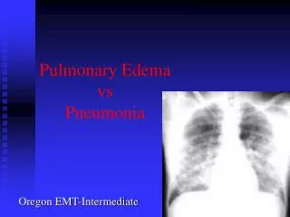
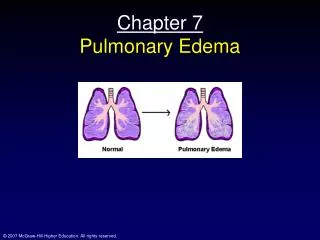
Acute Pulmonary Edema. The new england journal o f medicine. Clinical problem. cardiogenic pulmonary edema : hydrostatic or hemodynamic edema noncardiogenic pulmonary edema ; increased-permeability pulmonary edema, acute lung injury, or acute respiratory distress syndrome.

E N D
Acute Pulmonary Edema The new england journal o f medicine
Clinical problem • cardiogenic pulmonary edema : hydrostatic or hemodynamic edema • noncardiogenic pulmonary edema ; increased-permeability pulmonary edema, acute lung injury, or acute respiratory distress syndrome. • similar clinical manifestations.
cardiogenic pulmonary edema : diuretics and afterload reduction, underlying : CAD coronary revascularization. • noncardiogenic pulmonary edema : lung-protective strategy MV setting: low tidal volume (6 ml per kilogram of predicted body weight) and a plateau airway pressure less than 30 cm H2O reduces mortality in patients with acute lung injury. • severe sepsis, recombinant activated protein C4 and low-dose hydrocortisone should be considered • 找原因很重要, 怎麼找?
Mechanism: normal lung • Fluid and protein leakage (small gaps between capillary endothelial cells)alveolar interstitial space ( do not enter the alveoli because the alveolar epithelium is composed of very tight junctions) proximally into the peribronchovascular spacelymphatics remove most of this filtered • Movement of larger plasma proteins is restricted • hydrostatic force for fluid filtration across the lung microcirculation is approximately equal to the hydrostatic pressure in the pulmonary capillaries, partially offset by a protein osmotic pressure gradient
Mechanism: cardiogenic • rapid increase hydrostatic pressure increased transvascular fluid filtration • elevated pulmonary venous pressure from increased left ventricular end-diastolic pressure and left atrial pressure. • Mild elevations of left atrial pressure (18 to 25 mm Hg) cause edema in the perimicrovascular and peribronchovascular interstitial spaces. • Left atrial pressure rises further (>25 mm Hg), edema fluid breaks through the lung epithelium, flooding the alveoli with protein-poor fluid
Mechanism: noncardiogenic • increase in the vascular permeability of the lung, • increased flux of fluid and protein • Noncardiogenic pulmonary edema : high protein content • The net quantity of accumulated pulmonary edema : balance between the rate is filtered into the lung and the rate removed from the air spaces and lung interstitium.
evaluation • History and Physical Examination • Laboratory Testing • Chest Radiography • Echocardiography • Pulmonary-Artery Catheterization • stepwise approach
History and Physical Examination • All cause dyspnea and tachypnea. • Alveolar flooding arterial hypoxemia , cough and expectoration of frothy edema fluid. • history focus : underlying clinical disorder. • Cardiogenic : ischemia with or without MI; exacerbation of chronic systolic or diastolic HF; and dysfunction of the MV or AV; Volume overload. silent myocardial infarction or occult diastolic dysfunction may also manifest as acute pulmonary edema
History of noncardiogenic pulmonary edema • pneumonia, sepsis, aspiration of gastric contents, and major trauma associated with the administration of multiple blood-product transfusions. • S/S of infection, a decrease in the level of consciousness ( vomiting) , trauma, and details of medications and ingestions • 有時很難分的: AMI (cardiogenic edema) + syncope or cardiac arrest aspiration of gastric contents noncardiogenic edema mixed!!! • severe trauma or infection (noncardiogenic)fluid resuscitation volume overload increase in lung vascular hydrostatic pressure 真的很難分的….
PE • S3 gallop is relatively specific for elevated LVEDP and left ventricular dysfunction specificity : high (90 to 97 percent), but its sensitivity is low (9 to 51 percent). 因為病人使用呼吸器,會被干擾. • 其他心因性肺水腫PE的證據效力資料: 闕如 • A murmur consistent with valvular stenosis or regurgitation cardiogenic edema?? • The lung examination is not helpful.
PE • Elevated neck veins, an enlarged and tender liver, and peripheral edema suggest elevated central venous pressure. However, assessment of central venous pressure by physical examination in a critically ill patient can be difficult. • peripheral edema : not specific for left HF, other cause: hepatic or renal insufficiency, right heart failure, or systemic infection. • 用呼吸器,無法訴說腹部不適,要記的檢查PPU, infection等 • Noncardiogenic 四肢溫的, 即使沒有sepsis, cardiogenic: 冰的
Laboratory Testing • EKG :MI or ischemia. • Elevated troponin levels : damage to myocytes ; with severe sepsis in the absence of evidence for an acute coronary syndrome. • pulmonary edema of an unknown cause measurement of electrolytes, the serum OSM, and a toxicology screen may lead to the diagnosis of an unsuspected ingestion. • amylase and lipase acute pancreatitis
brain natriuretic peptide (BNP) • secreted predominantly by the cardiac ventricles in response to wall stretch or increased intracardiac pressures. • CHFplasma BNP levels correlate with LVEDP and PAWP. • BNP < 100 pg per milliliter indicates that HF is unlikely (negative predictive value, >90 percent), whereas a BNP > 500 pg per milliliter indicates that HF is likely (positive predictive value, >90 percent). • Between 100 and 500 pg per milliliter: inadequate diagnostic discrimination. • BNP levels can be elevated in critically ill patients even in the absence of heart failure. 24,25 Levels between 100 and 500 pg per milliliter are common in these patients • In one report, all eight patients with sepsis with normal left ventricular function had BNP levels above 500 pg per milliliter.
BNP is most useful in critically ill patients if the level is below 100 pg per milliliter. • BNP levels are also higher in patients with renal failure independent of heart failure, and a cutoff of below 200 pg per milliliter has been suggested to exclude heart failure when the estimated GFR < 60 ml/min. • BNP can also be secreted by the RV , and moderate elevations have been reported in patients with acute pulmonary embolism, cor pulmonale, and pulmonary hypertension.
Chest Radiography • Edema may not be visible until the amount of lung water increases by 30 percent. • any radiolucent material : alveolar hemorrhage, pus, and bronchoalveolar carcinoma will produce a radiographic image similar to that of pulmonary edema. • Technical issues can also reduce the sensitivity and specificity : rotation, inspiration, positive-pressure ventilation, position of the patient, and underpenetration or overpenetration • interobserver variability in the interpretation of radiographs.
Echocardiography • 49 critically ill patients with unexplained pulmonary edema or hypotension: use of 2-D transthoracic echocardiography and data generated from a pulmonary-artery catheter were in agreement in 86 percent of patients. • transthoracic echocardiography: first approach in patients in whom the history, physical and laboratory examinations, and the chest radiograph do not establish the cause of pulmonary edema. • less sensitive in identifying diastolic dysfunction normal result didn’t r/o
Pulmonary-Artery Catheterization • Pulmonary-artery catheterization: gold standard for determining the cause of acute pulmonary edema. • Other monitor: cardiac filling pressures, cardiac output, and systemic vascular resistanc. • pulmonary-artery occlusion pressure >18 mm Hg cardiogenic pulmonary edema or pulmonary edema due to volume overload. • rate of adverse advents was 4.5 to 9.5 percent • Common complications:hematoma at the insertion site, arterial puncture, bleeding, arrhythmias, and bloodstream infection; there were no fatalities.
Pulmonary-Artery Catheterization • Measurement of CVP should not be considered a valid substitute for PCWP, • there is often a poor correlation between the two. • Elevated CVP may reflect acute or chronic pulmonary arterial hypertension and right ventricular overload in the absence of any increase in left atrial pressure.
stepwise approach • noninvasive approaches for diagnosis will make mistakerepeated and ongoing assessment is necessary • 10 % of patients with acute pulmonary edema have multiple causes of edema. • 下列流程圖為作者臨床經驗所得
In one study that compared pulmonary-artery catheterization with clinical assessment by physicians: catheterization was superior. • However, that study predated the routine use of BNP and echocardiography, both of which are likely to increase the sensitivity and specificity of the noninvasive determination of the cause of pulmonary edema.