Download
1 / 70
700 likes | 729 Vues
Acute Interstitial Nephropathies (AIN) are renal disorders characterized by interstitial lesions leading to tubular injury, often caused by drugs, infections, or immune disorders. Learn about AIN epidemiology, etiology, diagnosis, and pathology.

E N D
Interstitial Nephropathies Definition IN are renal disorders, in which the main lesions are located in the renal interstitium which contributes to tubular injury later on, however with preserved glomerular integrity and intrarenal vascularization. Classification by evolution: -acute IN - chronic IN
Acute Interstitial Nephropathies (AIN) - Definition These are acute renal disorders, that usually occur in: a healthy kidney initially involving the interstitium. Rarely may occur on preexisting injuries of the renal parenchyma (either glomerular or vascular)
AIN Epidemiology AIN incidence is increasing, particularly due to exposure to a larger number of nephrotoxic agents (drugs, industrial toxic substances); AIN represents one of the main causes of acute renal failure (ARF), usually being reversible; In patients with ARF of unknown cause, subject to renal biopsy, AIN rate reaches 15%; AIN occurs at any age, with a maximal prevalence in the 50–60 decade.
AIN etiology Drug hypersensitivity Infections - acute pyelonephritis (the infectious agent is identified in the renal interstitium) - systemic infection – bacterial, viral, parasite, yeasts Immunologic disorders (SLE, Goodpasture's syndrome, acute renal transplante rejection, Sjogren’s syndrome, sarcoidosis) Unknown cause (idiopathic AIN)
AIN etiologic types Drug-induced AIN Infectious AIN Immunologic AIN Idiopathic AIN
Disease Processes Associated with AIN AIN of other etiology
AIN pathogenesis Immune - Following exposure to drugs (drug-induced AIN) - Within some immunologic disorders (immunologic AIN) - Within some systemic infections Infectious - acute bacterial pyelonephritis
AIN pathogenesis The precise disease mechanism is unclear, but antigen-driven immunopathology is the key mechanism. The presence of helper-inducer and suppressor-cytotoxic T lymphocytes in the inflammatory infiltrate suggests that T-cell mediated hypersensitivity reactions and cytotoxic T-cell injury are involved in pathogenesis of AIN. In some cases, humoral mechanisms are involved with complement proteins, immunoglobulins, and anti-TBM antibodies found in the interstitium.
AIN diagnosis Acute renal failure (ARF)with: Olyguria or normal diuresis; Malaise; Anorexia; Nausea and vomiting Clinical triad: fever, rash and arthralgia with acute onset is met in 1/3 of drug-induced AIN; with acute or subacute onset
AIN diagnosis • Eventually, bilateral low back pain (renal capsule distention); • Clinical evocative context for drug induced AIN (usually, after recently taking for 3 to 21 days a drug that has been shown to induce AIN).
AIN diagnosis Asymptomatic elevation in creatinine or blood urea nitrogen (BUN) or abnormal urinary sediment. Generalized hypersensitivity syndrome with fever, rash, eosinophilia, and oliguric renal failure. Clinical presentation variability Positive urinary sediment: leukocyturia, leukocyte cylinders, eventually bacteriuria (in infectious AINs)
AIN diagnosis Urinalysis examination shows: leukocyturia associated with leukocyte cylinders hematuria inferior to leukocyturia, mild to moderate proteinuria (tubular), eosinophiluria (non-specific, may be seen in prostatitis, vesical carcinoma as well). Proteinuria /24 hours does not exceed 1g, with predominating beta-2-microglobulin (tubular injury marker). In serum there is an elevation of: eosinophils count, and IgE byproducts of nitrogen retention (BUN, creatinine and uric acid).
AIN diagnosis Renal ultrasound exam does not show any specific for AIN imagistic data, however it allows to rule out the obstructive causes of ARI. It shows normal sized or enlarged kidneys, with increased echogenicity, and unclear boundaries between cortical and medullar layers Renal galium 67 scintigram allows for the differentiation of AIN (the kidneys capture the radiotracer) from acute tubular necrosis (absence of galium 67 capture at renal level).
Renal biopsy in AIN Considered to be the „golden standard” for AIN diagnosis, however rarely applied Indications for renal biopsy in ARF probably caused by AIN : - certain drug-induced AIN with mild ARF in which after 10 days after discontinuation of the incriminated drug, the evolution is unfavorable (ARI worsening); - in all patients with drug-induced AIN and ARF requiring acute hemodialysis, to rule out other causes of ARI and assess the long-term evolution.
AIN Pathology • The hallmark of AIN is the infiltration of inflammatory cells within the renal interstitium with associated edema, sparing the glomeruli and blood vessels. • Interstitial fibrosis is initially sparse, but develops later in the course of the illness. • Fibrotic lesions may be diffuse or patchy, beginning deep in the renal cortex, most prominently at the medullocortical junction.
AIN Pathology • The inflammatory infiltrate is typically composed of mononuclear cells and T lymphocytes, with a variable number of plasma cells and eosinophils. • Eosinophils may be totally absent from the infiltrate or may concentrate in small foci, forming eosinophilic microabscesses. • NSAIDs are commonly associated with glomerular involvement producing a minimal change disease. • In chronic interstitial nephritis, the cellular infiltrate is largely replaced by interstitial fibrosis.
AIN Pathology • Peritubular infiltration and occasional invasion of lymphocytes beneath the tubular basement membrane may occur with mild to extensive tubular damage, which may be difficult to distinguish from acute tubular necrosis (ATN). • Glomerular and renal vascular injuries are absent (except for collagenoses and vasculitides). • A third pathologic category is granuloma formation with epithelioid giant cells usually found in AIN secondary to tuberculosis, sarcoidosis, or Wegener's granulomatosis.
Tubulo-interstitial nephritis Inflammatory tubulo-interstitial condition, characterized by disorder of the concentration function and sometimes, kidney filtration function. There may be glomerular changes, however of a secondary character (as a rule).
TIN. Causes 1. Environmental factors • Cadmium • Lead • Ionizing radiation • Metabolic disorders • Hyperuricemia • Intrarenal artery embolism with cholesterol crystals • Systemic conditions • SLE • Sarcoidosis • Sjögren’s syndrome • HBV and HCV infection
TIN. Causes 4. Infections and invasions • bacterial • viral • parasitic 5. Tumours/hematologic disorders • Sickle cell anemia • Multiple myeloma • Light chain disease • Lymphoproliferative conditions 6. Hereditary • Hereditary tubulo-interstitial nephritis with cardiomegaly
TIN. Pathogenesis The vulnerability of tubulo-interstitial renal structures is caused mainly by the modest blood alimentation of this area, which is associated with a higher risk for ischemia and subsequent development of fibrosis. The main mechanism in case of renal disorder induced by NSAIDs is the insufficiency of local renal vaso-dilatator system (decrease of renal prostaglandins, due to their inhibition) In the development of analgesic nephritis an important role belongs to the total dose of the preparation and the duration of use, which sometimes may exceed 20-30 years.
TIN. Diagnosis Complete blood count: anemia, in case of drug-induced cause – with eosinophilia. Urinalysis: hyposthenuria, basic reaction, erytrhocyturia, sterile leukocyturia, tubular proteinuria. Biochemistry: elevated creatinine, potassium, and uric acid Zimnitki’s test: hyposthenuria, predomination of nighttime versus daytime diuresis.
TIN. Diagnosis Kidney USG : enlarged swollen kidneys (in acute phase) or shrunken, with uneven contour, cysts and calcinates (in chronic TINs) Kidney CT: shows the size of the kidneys, size of the cysts, and thickness of the cortical layer.
Supportive Care Measures AIN and TIN • Fluid and electrolyte management • Maintain adequate hydration • Avoid volume depletion or overload • Identify and correct electrolyte abnormalities • Symptomatic relief for fever and systemic symptoms • Symptomatic relief for rash • Avoid use of nephrotoxic drugs • Avoid use of drugs that impair renal blood flow • Adjust drug dosages for existing level of renal function
Infectious AIN Acute pyelonephritis (APN) AIN secondary to a systemic infection
Acute pyelonephritis (APN) Definition: acute nephropathy characterized by an infection of the interstitial tissue and renal pelvis. APN classification depending on the presence of favoring factors: ▪ uncomplicated APN (without any favoring factors); ▪ complicated APN – with favoring factors
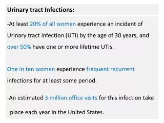
Causes • E.coli is the cause of 80–85% of urinary tract infections. • Staphylococcus saprophyticus being the cause in 5–10%. • Rarely they may be due to viral or fungal infections. • Other bacterial causes include: Klebsiela, Proteus, Pseudomonas and Enterobacter. These are uncommon and typically related to abnormalities of the urinary system or urinary catheterisation. • UTI due to Staphylococcus aureus typically occur secondary to blood-borne infections.
APN symtoms ▪ sudden onset within hours to 1-2 days; ▪ infectious syndrome (fever, chills, sweating, headache, myalgia, arthralgia, dizziness, nausea, vomiting); ▪ deaf low-back pain (uni- or bilateral) or, randomly, cramping; ▪ cystitis syndrome (burning at urination, pollakiuria, dysuria, cloudy urine) ;
Physical exam in APN At palpation: - lumbar sensitivity, - painful ureteral points, - painful costo-muscular and costo-vertebral points; Percussion of the lumbar area: positive Giordano maneuver; Cardiovascular symptoms: tachicardia corresponding to fever, normal either slightly decreased BP; Dehydration signs (induced by fever): dry tongue, persistent skin fold.
APN types Serous Aposthematous – an acute purulent process associated with formation of multiple pustules (aposthemas), particularly in cortex Renal carbuncle – the inflammatory process develops in the cortical layer of the kidney in a limited area and is characterized by a combination of ischemic, necrotic process and pus. Renal abscess – occurs in severe inflammation of renal parenchyma due to melting of suppurative parenchyma, the merging of aposthemas, either destruction of a renal carbuncle.
APN. Differentials Acute pancreatitis Acute cholecystitis Acute appendicitis Acute salpingitis Acute prostatitis Various acute infections – in the elderly
APN. Diagnosis • Complete blood count • Leukocytosis with left shift in white blood cell counts • Elevated ESR
APN. Diagnosis • Urinalysis : • Leukocyturia – more than 6 in the v/f • Proteinuria – usually false, due to degradation of leucocytes, either tubular, cause by tubulopathy; does not exceed 1gr/24h, more frequently varying from 0.033 up to 1.0. • Erythrocyturia – in obstructive uropathies. • Pyuria – leukocyturia + bacteriuria • In case of urine contamination with genital discharge one can see leukocytes in groups, flat epithelium and mucus in large amounts.
APN. Diagnosis • Biochemical blood assessment: • Azotemia (elevated blood urea nitrogen, creatinine, uric acid) – is not a predictive sign of renal failure because of the localized character of inflammation. • It may be significant in case of total obstruction of the upper urinary tract with a stone or in case of an acute pathologic process in the contralateral kidney.
APN. Diagnosis • Urine culture – shows growth of bactrial colonies, however it may be sterile when on antibiotic therapy. • Neciporenco’s test: • Leukocytes – up to 4000 per ml of urine • Erythrocytes – up to 2000 per ml of urine • Cylinders – up to 20 per ml of urine
APN. Diagnosis Ultrasoud picture of bilateral pyelonephritis. On sections through areas of gate of both kidneys moderately enlarged kidney sinuses with the expressed thickening and multiple layers of walls are visualized. The kidneys size is increased due to parenchyma, its ultrasound density last is raised.
APN. Diagnosis Intravenous urography. Retrograde pyelography – reflux from the upper calyx.
APN. Complications Chronic renal failure Acute renal failure Necrotizing papillitis Paranephritis Urosepsis Kidney stones
APN. Evolution Complete recovery Chronic disease In case of a unilateral purulent injury – shrinkage with function loss
Chronic Pyelonephritis Definition – infection-induced chronic inflammation of the renal interstitium and pyelocaliceal system. Etiology – the same as in APN
CPN Pathogenesis Disorders of the normal urodinamics favor the appearance of inflammation by infection of the stalled urine. The increase in the pressure in the pyelocaliceal system leads to compression of the fornical veins with their rupture, which favors the direct penetration of infection from pelvis to the renal microcirculation, causing secondary hematogenous infection of the renal cortex and interstitium.
CPN pathogenesis A frequent cause of non-obstructive CPN is the vesical-ureteral reflux, which in time leads to kidney shrinkage. When in the shrunken kidney perivascular sclerosis predominates, the clinical picture of progressive hypertension appears, which is less prominent in case of periglomerular and peritubular sclerosis.