Download
1 / 23
260 likes | 1.08k Vues
Ventilation: the Mechanics of Breathing. The way mammals ventilate their lungs. Organs of the Respiratory system. Lungs close up…. Bronchial Tree Consisting of the Passageways that Connect the Trachea and Alveoli . Breathing.

E N D
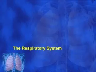
Ventilation: the Mechanics of Breathing The way mammals ventilate their lungs
Lungs close up… • Bronchial Tree Consisting of the Passageways that Connect the Trachea and Alveoli
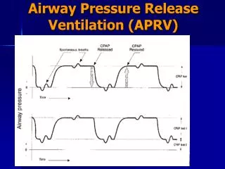
Breathing • The movement of air into and out of the lungs (ventilation) results from a pressure difference between the thoracic cavity and the atmosphere. • This pressure difference is created by changing the volume of the thoracic cavity as shown in the animation below.
Involuntary Respiration. The basic rhythm of breathing occurs without conscious effort. The inspiratory center located in the medulla sets the basic rhythm by automatically initiating inspiration with a two second burst of nerve impulses to the diaphragm and the external intercostal muscles. Contraction of the diaphragm and the external intercostal muscles draws air into the lungs. Involuntary Respiration
The Expiratory Center. The expiratory center is located in the medulla. This center functions during forced expiration stimulating the internal intercostal and abdominal muscles to contract.
Inhalation • During inhalation, the diaphragm contracts and flattens and the external intercostal muscles draw the ribs upward and outward. • This increase in thoracic volume results in a decrease in intrapulmonary pressure. • Air enters the lungs to stabilize the pressure difference between the external atmosphere and the internal compartments of the lungs. • Normal inhalation is an active process, requiring muscular work.
During quiet breathing, intercostals maintain the rigidity of the chest wall. Otherwise, reduced intra-thoracic pressure would cause the chest wall to collapse inwards. External Intercostals (on the outside of the ribcase) wrap around from the back of the rib almost to the end of the bony part of the rib in front. . They elevate the ribs. Internal Intercostals in the inside of the ribcase) extend from the front of the ribs, and go around back, past the bend in the ribs . They depress the ribs.
Expiration Animation: Alveolar Pressure Changes During Inspiration and Expiration • Exhalation is normally a passive process. • The diaphragm and external intercostal muscles relax decreasing the volume of the thoracic cavity. • This causes the pressure within the lungs to exceed the atmospheric pressure. • Air is expelled from the lungs.
Biology http://www.bishopstopford.com/faculties/science/arthur/Breathing%20System%20Drag%20%26%20Drop.swf
Forced Exhalation • During a forced exhalation, the internal intercostal muscles contract, depressing the rib cage. • The abdominal muscles contract, pushing the organs in the abdominal cavity against the diaphragm. • The thoracic volume decreases to a level lower than achieved in normal exhalation. • These muscles are used to counteract the effects of obstructive pulmonary disorders.
ERV • These muscles are used during a forced exhalation to determine the expiratory reserve volume (ERV). • ERV is - the maximum volume of gas that can be forcefully exhaled after a normal exhalation (tidal volume).
The volume of the lungs is divided into four functional compartments, lung volumes. Combinations of two or more lung volumes are called a lung capacity. BTPS- Body Temperature, ambient Pressure and Saturated with water vapor. Standard conditions for calculating lung volumes. Other Terms of Breathing
Terms • tidal volume ( TV ) - the volume of gas inspired or expired during each normal (unforced) ventilation cycle (volume of air moved into the lungs in a single breath. • inspiratory reserve volume ( IRV ) -the maximum amount of gas that can be forcefully inhaled after a normal inhalation. • expiratory reserve volume ( ERV ) - the maximum volume of gas that can be forcefully exhaled after a normal exhalation.
Terms • residual volume ( RV ) - the amount of gas left in the lungs after a maximum (forced) exhalation. Necessary otherwise the lungs would collapse. • total lung capacity ( TLC ) - the amount of gas in the lungs after a maximum (forced) inhalation. TLC = IRV + TV + ERV + RV • vital capacity ( VC )-the maximum volume of gas that can be exhaled by voluntary effort after a maximum inhalation. VC = IRV + TV + ERV
Terms • inspiratory capacity ( IC ) - the maximum amount of gas that can be inhaled after a normal (unforced) exhalation. IC = IRV + TV • functional residual capacity ( FRC ) - the amount of gas left in the lungs after a normal (unforced) exhalation. FRC = ERV + RV
Terms • minute volume ( MV ) - or minute respiratory volume - the volume of air breathed per minute - MV = tidal volume x respiratory rate (normally 5-8 liters per minute). • Alveolar ventilation/minute ( VA )- is the portion of the minute volume of ventilation which reaches those areas of the lung concerned with gas exchange. VA = (Tidal volume minus Dead Space) x Rate. Averages about 3.5 to 5.0 liters per minute. Alveolar ventilation/minute is the best criterion for effectiveness of breathing.
Question?? • As Stanley sits in the drivers seat at rest, which muscles are contributing the majority of his breathing? 1. Internal intercostals2. External intercostals3. Accesory muscles4. Diaphragm5. Psoas major
4. Diaphragm • In a healthy adult, the diaphragm is the dominant muscle of respiration at rest • The diaphragm is a musculotendinous sheet separating the thorax from the abdomen. It is attached to the thoracic cage under the lower ribs.
Respiratory Control • Homeostasis: maintenance of a constant internal environment • Homeostasis in breathing involves the regulation of CO2 and O2 levels in the body. These gas levels are regulated by 2 control systems: • Nervous control: an area in the brain called the respiratory centre (medulla oblongata) has nerve fibres that connect it to the muscles of the rib cage and the diaphragm • Chemical control: chemical receptors in the walls of arteries detect changes in levels of CO2 and send signals to the respiratory centre. Breathing Mechanism explained using an Animation - Tutorvista.com
Factors affecting breathing rate • Breathing can be affected by internal body conditions and environmental conditions • Internal: • Increased levels of CO2 in the blood • Respiratory disease (common cold, influenza, TB) • Yawning • Amount of cellular activity (muscle cells require more energy when they are worked, therefore the amount of gas exchange will increase) b) Environmental: • At higher altitudes, the air is thinner and contains less O2, therefore breathing rate increases • Emotion • Dust, pollen • Smoke (carbon cannot be filtered) • Industrial chemicals (CO, ammonia, methane cause inflammation of respiratory tissue)
The Basal Metabolic Rate • B.M.R.: the rate at which energy is released by your body while at rest • The B.M.R. is found by measuring O2 intake and CO2 output. The B.M.R. is higher for males but decreases with age.