Download

1 / 1
10 likes | 142 Vues
Assessment of Microgroove-Aligned Endothelial Cells for in vitro Simulations. . Jennifer L. Fischer 1 , Lindsay N. Gray 1 , Christine A. Trinkle 2 , Richard E. Eitel 1 , Kimberly W. Anderson 1 Department of Chemical and Materials Engineering, University of Kentucky, Lexington 1

E N D
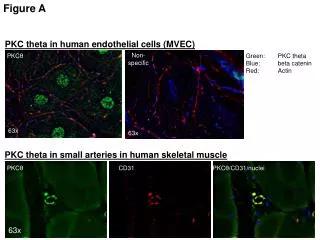
Assessment of Microgroove-Aligned Endothelial Cells for in vitro Simulations  Jennifer L. Fischer1, Lindsay N. Gray1, Christine A. Trinkle2, Richard E. Eitel1, Kimberly W. Anderson1Department of Chemical and Materials Engineering, University of Kentucky, Lexington1 Department of Mechanical Engineering, University of Kentucky, Lexington2 ABSTRACT MATERIALS AND METHODS RESULTS - MORPHOLOGY RESULTS - CHEMISTRY VCAM-1 Staining Micropatterning and Seeding Protocols F-actin Staining The circulatory system is lined with a thin layer of endothelial cells, which compose the lining of the vessel walls. Adhesion of cancer cells to this endothelial cell lining is an important step in the metastatic cascade and researchers are currently using in vitro techniques to investigate these interactions. Under static culture conditions, endothelial cells grow in a characteristic cobblestone pattern rather than growing in straight lines due to the absence of shear stresses that would normally be found in circulatory vessels. It has recently been suggested that such changes in cell morphology can affect surface expression profiles, which may alter how frequently or strongly cancer cells bind to endothelial cells. While flow adapting endothelial cells is important prior to studying cancer cell binding in vitro, traditional methods can be cumbersome due to the fact that the cells have to be exposed to flow for an extended period of time under controlled environmental conditions. In this study, we are investigating the efficacy of a microgroove-aligned flow adaptation methodfor acquiring a flow adapted phenotype. • Microgrooves were created on glass slides using the negative epoxy-based photoresist, SU-8 2000.5, as shown below. • Human Umbilical Vein Endothelial Cells (HUVECs) were then seeding onto unmodified (control) glass slides and micropatterned slides as shown below. Endothelial Cells: Cobblestone vs. Unidirectional Growth • F-actin staining was used to stain the actin fibers in the cellular cytoskeleton so that the aspect ratios and orientation angles could be tabulated more readily • Images show noticeable elongation of HUVECs on micropatterned versus control slides regardless of seeding density • Elongation and orientation were compared at high and low seeding densities for unmodified control and micropatterned slides • High density: 62,000cells/cm2, Low density: 25,000 cells/cm2 • Initial fluorescent images of antibody tagging to evaluate surface expression • Primary Antibody: Anti-VCAM-1 Antibody, clone P3C4 • Secondary Antibody: Goat Anti-Mouse IgG (Fc), Fluorescein Conjugated • Staining completed for N=3 slides for both the unmodified control and micropatterns. Unmodified slidesstimulated with 480U/ml TNF-α (tumor necrosis factor alpha) were also used as a positive control (N=3). Three images were analyzed per slide. Morphology and Surface Chemistry Evaluation Protocols • The HUVEC morphology was characterized by aspect ratios and orientation angles. • Aspect ratio = length of cell/width of cell • Orientation angle = deviation from horizontal • Grooves defined as 0° • Positive deviations represent above horizontal, negative represent below • The surface chemistry was first evaluated by examining vascular cell adhesion molecule VCAM-1 expression due to its significance in the metastatic cascade. This was done using the following protocol: Aspect Ratio Comparison Preliminary results from images and median pixel intensities (histogram peaks) suggest upregulation of VCAM-1 on the surface of microgroove-aligned HUVECs. This was not statistically significant in comparisons of mean pixel intensities. CONCLUSIONS • Using microgrooves created with SU-8 photoresist on glass, HUVECs were successfully cultured statically in a more elongated and unidirectional form reminiscent of in vivo morphology • Initial fluorescent images suggest upregulation of VCAM-1 on micropatterned glass slides which is more representative of the in vivo surface chemistry. The average histograms derived from the images also suggest upregulation (as evidenced by positive shift in median peak) on the microgrooves, however this effect was not apparent in the means likely due to the asymmetry from cutoffs of autofluorescence and nonspecific staining on the left and the variability in the right tail regions. RESULTS – APPROACH VALIDATION Atomic Force Microscopy Characterization OBJECTIVES • At both seeding densities, statistically higher aspect ratios were obtained for the micropatterned versus the control slides (p-value <0.05) • Error bars represent SE • In unmodified controls, n= 60. In micropatterned studies, n = 138 and n = 114 FUTURE WORK Grooves Glass • Examine surface expression of ICAM-1, E-selectin and P-selectin • Quantify results of surface chemistry in spectrophotometer for comparison • Compare results to cells flow adapted in a parallel plate flow chamber Overall Objective • The overall objective of this work was to create a static culture system that better mimics thechemistry and morphology of endothelial cells grown in vivo usingmicrofabrication techniques. Specific Objectives • Investigate and compare morphology of endothelial cells grown on micropatterned grooves with those grown on a blank glass slide by quantifying aspect ratios and orientation angles. • Investigate surface expression of cells grown on micropatterned grooves in comparison to cells grown on a blank slides using fluorescent tagging of antibodies for the following proteins involved in tumor cell adhesion to the endothelium: ICAM-1, VCAM-1, and E-selectin and P-selectin. Orientation Angle Comparison ACKNOWLEDGEMENTS • UKY NCI CNTC (Grant # R25CA153954) • UKY NSF IGERT Grant in Bioactive Interfaces and Devices (NSF Award # 0653710) • CeNSE Laboratory at the University of Kentucky • Support from the National Science Foundation REU Program #EEC-1156667 and the Bucks For Brains Program, Office of Undergraduate Research, University of Kentucky REFERENCES • Shown above are orientation angle histograms for the high density micropatterned slides (left) vs. control (right) • Micropatterned slides show small distribution mostly between ± 20° • Control slides show large distribution between ± 100° Results demonstrate elongated, unidirectional growth achieved • The AFM was used to confirm that our methods for patterning SU-8 2000.5 were correct and resulted in the desired topography. • Average groove depth was 500 nm as expected • Consistent trench widths and spacing was achieved • MicroChem. http://www.microchem.com/Prod-SU8_KMPR.htm (April 2012). • Cines et al.Endothelial Cells in Physiology and in the Pathophysiology of Vascular Disorders. Blood Journal. 1998; vol 91 • Park, T; Shuler M. Integration of Cell Culture and Microfabrication Technology. Biotechnol.Prog. 2003, 19, 243-253