Western Blot Analysis of sFRP1 and Associated Protein Interactions in Prostate Cancer Cell Lines
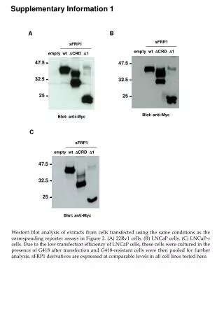
This study presents Western blot analyses of sFRP1 variations in 22Rv1 and LNCaP cell lines. Transfections were conducted under conditions consistent with reporter assays in Figure 2. Due to low transfection efficiency, LNCaP cells were treated with G418 to select resistant clones for analysis. The results show comparable expression levels of sFRP1 derivatives across tested cell lines. Additional analyses include interactions with Wnt proteins and beta-catenin, highlighting the complexity of sFRP1's role in cancer signaling pathways.

Western Blot Analysis of sFRP1 and Associated Protein Interactions in Prostate Cancer Cell Lines
E N D
Presentation Transcript
47.5 32.5 25 47.5 B 32.5 sFRP1 empty wt DCRD D1 25 47.5 32.5 25 Blot: anti-Myc C sFRP1 empty wt DCRD D1 Blot: anti-Myc Supplementary Information 1 A sFRP1 empty wt DCRD D1 Blot: anti-Myc Western blot analysis of extracts from cells transfected using the same conditions as the corresponding reporter assays in Figure 2. (A) 22Rv1 cells, (B) LNCaP cells, (C) LNCaP-r cells. Due to the low transfection efficiency of LNCaP cells, these cells were cultured in the presence of G418 after transfection and G418-resistant cells were then pooled for further analysis. sFRP1 derivatives are expressed at comparable levels in all cell lines tested here.
Supplementary Information 2 A - - + + sFRP1 - - + + Wnt3A Blot: anti-Myc (for sFRP1) B - - sFRP1 + + b-catenin WT SA WT SA Blot: anti-Myc (for sFRP1) IP: anti-HA Blot: anti-b-catenin C - - + + sFRP1 - - + + Wnt5A Blot: anti-Myc (for sFRP1) Blot: Wnt5A Western blot analysis of extracts from 22Rv1 cells transfected using the same conditions as the aforementioned reporter assays. (A) Transfections corresponding to Figure 3 (Wnt3a antibodies were not sensitive enough to detect the low level of transfected Wnt3a in these experiments), (B) Transfections corresponding to Figure 4, (C) Transfections corresponding to Figure 6D. Regarding (B), detection of HA-tagged beta-catenin in cell extracts was not possible. Therefore, anti-HA ips were probed for HA-tagged beta-catenin.