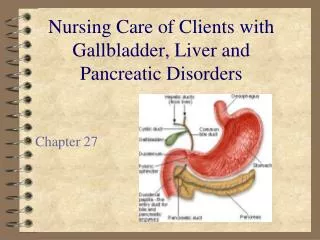
Gallbladder Disorders
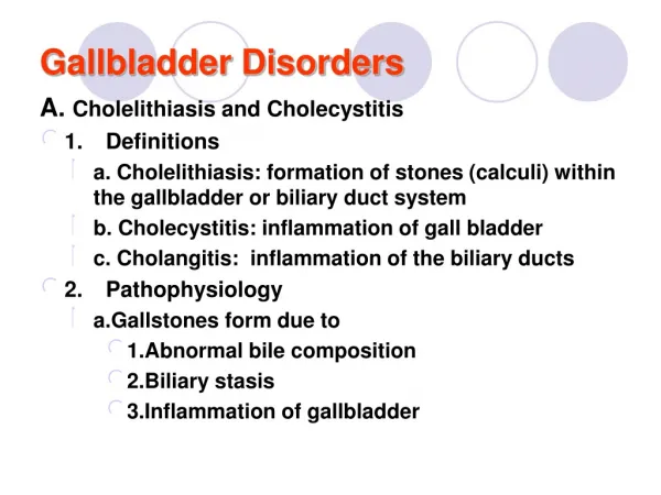
Gallbladder Disorders. A. Cholelithiasis and Cholecystitis 1. Definitions a. Cholelithiasis: formation of stones (calculi) within the gallbladder or biliary duct system b. Cholecystitis: inflammation of gall bladder c. Cholangitis: inflammation of the biliary ducts 2. Pathophysiology


Gallbladder Disorders
E N D
Presentation Transcript
Gallbladder Disorders A. Cholelithiasis and Cholecystitis • 1. Definitions • a. Cholelithiasis: formation of stones (calculi) within the gallbladder or biliary duct system • b. Cholecystitis: inflammation of gall bladder • c. Cholangitis: inflammation of the biliary ducts • 2. Pathophysiology • a.Gallstones form due to • 1.Abnormal bile composition • 2.Biliary stasis • 3.Inflammation of gallbladder
Gallbladder Disorders • b. Most gallstones are composed primarily of bile (80%); remainder are composed of a mixture of bile components • c. Excess cholesterol in bile is associated with obesity, high-cholesterol diet and drugs that lower cholesterol levels • d. If stones from gallbladder lodge in the cystic duct • 1. There can be reflux of bile into the gallbladder and liver • 2. Gallbladder has increased pressure leading to ischemia and inflammation • 3. Severe ischemia can lead to necrosis of the gall bladder • 4. If the common bile duct is obstructed, pancreatitis can develop
Gallbladder Disorders Risk factors for cholelithiasis • a. Age • b. Family history, also Native Americans and persons of northern European heritage • c. Obesity, hyperlipidemia • d. Females, use of oral contraceptives • e. Conditions which lead to biliary stasis: pregnancy, fasting, prolonged parenteral nutrition • f. Diseases including cirrhosis, ileal disease or resection, sickle-cell anemia, glucose intolerance
Gallbladder Disorders Manifestations of cholelithiasis • a. Many persons are asymptomatic • b. Early symptoms are epigastic fullness after meals or mild distress after eating a fatty meal • c. Biliary colic (if stone is blocking cystic or common bile duct): steady pain in epigastric or RUQ of abdomen lasting up to 5 hours with nausea and vomiting • d. Jaundice may occur if there is obstruction of common bile duct
Gallbladder Disorders Manifestations of acute cholecystitis • a. Episode of biliary colic involving RUQ pain radiating to back, right scapula, or shoulder; the pain may be aggravated by movement, or deep breathing and may last 12 – 18 hours • b. Anorexia, nausea, and vomiting • c. Fever with chills
Gallbladder Disorders Complications of cholecystitis • a. Chronic cholecystitis occurs after repeated attacks of acute cholecystitis; often asymptomatic • b. Empyema: collection of infected fluid within gallbladder • c. Gangrene of gall bladder with perforation leading to peritonitis, abscess formation • d. Pancreatitis, liver damage, intestinal obstruction
Gallbladder Disorders Collaborative Care • a. Treatment depends on the acuity of symptoms and client’s health status • b. Clients experiencing symptoms are usually treated with surgical removal of the stones and gallbladder Diagnostic Tests • a. Serum bilirubin: conjugated bilirubin is elevated with bile duct obstruction • b. CBC reveals elevation in the WBC as with infection and inflammation • c. Serum amylase and lipase are elevated, if obstruction of the common bile duct has caused pancreatitis • d. Ultrasound of gallbladder: identifies presence of gallstones • e. Other tests may include flat plate of the abdomen, oral cholecytogram, gall bladder scan
Gallbladder Disorders Treatment • a. Treatment of choice is laparoscopic cholecystectomy • b. If surgery is inappropriate due to client condition • 1. May attempt to dissolve the gallstones with medications • 2. Medications are costly, long duration • 3. Stones reoccur when treatment is stopped Laparoscopic cholecystectomy • a. Minimally invasive procedure with low risk of complications; required hospital stay< 24 hours. • b. Learning needs of client and family/caregiver include pain control, deep breathing, mobilization, incisional care and nutritional/fluids needs • c. Client is given phone contact for problems
Gallbladder Disorders Some clients require a surgical laparotomy (incision inside the abdomen) to remove gall bladder • a. client will have nasogastric tube in place post-operatively and require several days of hospitalization • b. If exploration of the common bile duct is done with the cholecystectomy, the client may have a T-tube inserted which promotes bile passage to the outside as area heals Clients with cholelithiasis and cholecystitis prior to surgery can avoid future attacks by limiting fat intake Nursing Diagnoses • a. Pain • b. Imbalanced Nutrition: Less than body requirements • c. Risk for Infection
Liver Disorders A. Hepatitis • 1. Definition: inflammation of the liver due to virus, exposure to alcohol, drugs, toxins; may be acute or chronic in nature • 2. Pathophysiology: metabolic functions and bile elimination functions of the liver are disrupted by the inflammation of the liver.
Liver Disorders Viral Hepatitis • 1. Types (causative agents) a. Hepatitis A virus (HAV) Infectious hepatitis • 1. Transmission: fecal-oral route, often contaminated foods, water or direct contact, blood transfusions, contaminated equipment • 2. Contagious through stool up to 2 weeks before symptoms occur; abrupt onset • 3. Benign, self limited; symptoms last up to 2 months
Liver Disorders • Prevention of Hepatitis A • Good handwashing • Good personal hygiene • Control and screening of food handlers • Passive immunization • Incubation period :20-50 days (short incubation period)
Liver Disorders • Incidence • More common in fall and winter months • Usually found in children and young adults • Infectious for 3 weeks prior and 1 week after developing jaundice • Clinical recovery 3-16 weeks
Liver Disorders Hepatitis B virus (HBV) • 1. Transmission: • infected blood and body fluids, • parenteral route with infusion • ingestion or inhalation of the blood of an infected person • Contaminated needles, syringes, dental instruments • Oral or sexual contact • High risk individuals include homosexual, IV drug abusers, persons with multiple sexual partners, medical workers • 2. Liver cells damaged by immune response; increased risk for primary liver cancer; causes acute and chronic hepatitis, fulminant hepatitis and carrier state
Liver Disorders • Hepatitis B • Prevention • Screen blood donors • Immunization
Liver Disorders Hepatitis C virus (HCV) • 1. Transmission: infected blood and body fluids; injection drug use is primary factor • 2. Initial manifestations are mild, nonspecific • 3. Primary worldwide cause of chronic hepatitis, cirrhosis, liver cancer • 4. Usual incubation period 7-8 weeks
Liver Disorders Hepatits B-associated delta virus (HDV) • 1. Transmission: infected blood and body fluids; causes infection in people who are also infected with hepatitis B • 2. Causes acute or chronic infection • Hepatitis D • Transmitted through oral-fecal contaminated water, course of illness resembles hepatitis A
Liver Disorders Hepatitis E virus (HEV) • 1. Transmission: fecal-oral route, contaminated water supplies in developing nations; rare in U.S. • 2. Affects young adults; fulminant in pregnant women
Liver Disorders Disease Pattern Associated with hepatitis (all types) • A .Incubation Phase (period after exposure to virus): no symptoms • B Prodromal Phase (preicteric – before jaundice) • 1. “Flu” symptoms: general malaise, anorexia, fatigue, muscle and body aches • 2. Nausea, vomiting, diarrhea, constipation, and mild RUQ abdominal pain • 3. Chills and fever • c.Icteric (jaundiced) Phase • 1 5 – 10 days after prodromal symptoms • 2. Jaundice of the sclera, skin and mucous membranes occurs • 3. Elevation of serum bilirubin • 4. Pruritis • 5. Stool become light brown or clay colored • 6. Urine is brownish colored
Liver Disorders Convalescent Phase • 1. In uncomplicated cases, symptoms improve and spontaneous recovery occurs within 2 weeks of jaundice • 2. Lasts several weeks; continued improvement and liver enzymes improve
Liver Disorders Chronic Hepatitis • a. Chronic hepatitis: chronic infection from viruses: HBV, HBC, HBD • 1. Few symptoms (fatigue, malaise, hepatomegaly) • 2. Primary cause of cirrhosis, liver, cancer, liver transplants • 3. Liver enzymes are elevated • b. Fulminant hepatitis; rapidly progressive disease with liver failure developing within 2 – 3 week of onset of symptoms; rare, but usually due to HBV with HBD infections • c. Toxic hepatitis • 1. Hepatocellular damage results from toxic substances • 2. Includes alcoholic hepatitis, acute toxic reaction or chronic use
Liver Disorders Collaborative Care: Focus is on determination of cause, treatment and support, and prevention future liver damage Diagnostic Tests a. Liver function tests • 1. Alanine aminotransferase (ALT): specific to liver • 2. Aspartate aminotransferase (AST): heart and liver cells • 3. Alkaline phosphatase (ALP): liver and bone cells • 4. Gamma-glutamyltransferase (GGT): present in cell membranes; rises with hepatitis and obstructive biliary disease • 5. Lactic dehydrogenase (LDH): present in many body tissues; isoenzyme, LDH5 is specific to the liver • 6. Serum bilirubin levels: total, conjugated, unconjugated
Liver Disorders b. Lab tests for viral antigens and antibodies associated with types of viral hepatitis c. Liver biopsy: tissue examined to detect changes and make diagnosis • 1. Preparation: signed consent; NPO 4 – 6 hours before • 2. Prothrombin time and platelet count results; may need Vitamin K first to correct • 3. Client voids prior to procedure, supine position • 4. Local anesthetic; client instructed to hold breath during needle insertion • 5. Direct pressure applied to site after sample obtained; client placed on right side to maintain site pressure • 6. Vital signs monitored frequently for 2 hours • 7. No coughing, lifting, straining 1 – 2 weeks afterward
Liver Disorders Medications for prevention of hepatitis • a. Vaccines available for Hepatitis A and B • b. Vaccine for Hepatitis B recommended for high-risk groups • c. Post exposure prophylaxis recommended for household and sexual contacts of persons with HAV or HBV • d. Hepatitis A prophylaxis: single dose of immune globulin within 2 weeks of exposure • e. Hepatitis B prophylaxis: Hepatitis B immune globulin (HBIG) for short-term immunity; HBV vaccine may be given at the same time
Liver Disorders Treatment • a. Medications • 1. Medication for acute hepatitis C: interferon alpha to prevent chronic hepatitis • 2. Chronic Hepatitis B: interferon alpha intramuscular or subcutaneously or lamivudine • 3. Chronic Hepatitis C: interferon alpha with ribavirin (Rebetol) oral antiviral drug
Liver Disorders b. Acute hepatitis treatment • 1. As needed bedrest • 2. Adequate nutrition • 3. Avoid substances toxic to the liver especially alcohol c. Complementary therapies: Milk thistle (silymarin) 8. Nursing Care: Teaching about prevention by stressing • a. Hygiene • b. Handwashing, especially for food handlers • c. Blood and body fluids precautions • d. Vaccines for persons at high risk • e. Restrict use of alcohol • f. Abstain from sexual activity during communicable period
Liver Disorders Nursing Diagnoses • a. Risk for Infection • 1.Standard precautions, proper hand washing at all times • 2.Reporting of contagious disease to health department to control spread of disease • b. Fatigue • 1.Scheduling planned rest periods • 2.Gradual increase of activity with improvement • c. Imbalanced Nutrition: Less than body requirements • 1.High caloric diet with adequate carbohydrates • 2.Small frequent meals; nutritional supplements • d. Body Image Disturbance Home care must include proper infection control measures; continuing medical care
Cirrhosis Definition • a. End state of chronic liver disease • b. Progressive and irreversible • c. Tenth leading cause of death in U.S. Pathophysiology • a. Functional liver tissue gradually destroyed and replaced with fibrous scar tissue • b. As hepatocytes are destroyed, metabolic functions are lost • c. Blood and bile flow within liver is disrupted • d. Portal hypertension develops • Portal vein receives blood from the intestines and spleen, so as portal hypertension increases the blood flows back in the esophageal and umbilical veins causing ascites as well as splenomegaly
Cirrhosis Alcoholic cirrhosis (Laennec’s cirrhosis) • a. Alcohol causes metabolic changes in liver leading to fatty infiltration (stage in which abstinence from alcohol could allow liver to heal) • b. With continued alcohol abuse, inflammatory cells infiltrate liver causing necrosis, fibrosis and destruction of liver tissue • c. Regenerative nodules form, liver shrinks and is nodular • d. Malnutrition commonly present
Cirrhosis Biliary cirrhosis: Bile flow is obstructed and is retained within liver causing inflammation, fibrosis and regenerative nodules to form • increased skin pigmentation resembling a deep tan, jaundice and pruritus Posthepatic cirrhosis: Chronic hepatitis B or C and unknown cause leads to liver shrinkage and nodule formation with extensive liver cell loss and fibrosis
Cirrhosis • Cardiac cirrhosis • Right sided CHF. Liver is swollen, yet reversible if CHF is treated • Nonspecific, metabolic cirrhosis • Metabolic problems, infectious disease, infiltrative disease, GI disease could be the cause
Cirrhosis Manifestations • a. Early: liver enlargement and tenderness, dull ache in RUQ, weight loss, weakness, fatigue, anorexia, diarrhea or constipation • b. Progresses to impaired metabolism causing bleeding, ascites, gynecomastia in men, infertility in women, jaundice, neurological changes, ascites, peripheral edema, anemia, low WBC and platelets
Cirrhosis Complications • a. Portal hypertension: • shunting of blood to collateral blood vessels leading to engorged veins in esophagus, rectum and abdomen, ascites • Pressures within the portal venous system become elevated as liver damage obstructs the free flow of blood through the organ • b.Splenomegaly: anemia, leucopenia, thrombocytopenia
Cirrhosis • c.Ascites: • accumulation of abdominal fluid rich in protein; hypoalbuminemia, sodium and water retention • Result of portal hypertension • Increased level of aldosterone
Cirrhosis • d. Esophageal varices: thin walled dilated veins in esophagus which may rupture leading to massive hemorrhage • Secondary to portal hypertension • Bleeding may occur as a result of mechanical trauma, ingestion of coarse food
Cirrhosis • e. Hepatic encephalopathy: from accumulated neurotoxins in blood; ammonia produced in gut is not converted to urea which is normally excreted and accumulates in blood and is trapped in the brain; medications may not be metabolized and add to mental changes including personality changes, slowed mentation, asterixis (liver flap); progressing to confusion, disorientation and coma
f. Hepatorenal syndrome: renal failure with azotemia • Anorexia • Fatigue • Weakness • Fluid retention leads to hyponatremia and fluid overload • Needs hemodialysis for hyperkalemia and fluid overload
Cirrhosis Collaborative Care: Holistic care to client and family addressing physiologic, psychosocial, spiritual needs Diagnostic Tests • a. Liver function tests (ALT, AST, alkaline phosphatase, GGT); elevated, but not as high as with acute hepatitis • b. CBC and platelets: anemia, leucopenia, thrombocytopenia • c. Prothrombin time: prolonged (impaired coagulation due to lack of Vitamin K) • d. Serum electrolytes: deficiencies in sodium, potassium, phosphate, magnesium • e. Bilirubin: elevated failing liver can’t bind bilirubin • f. Serum albumin: hypoalbuminemia • g. Serum ammonia: elevated • h. Serum glucose and cholesterol
Cirrhosis • i. Abdominal ultrasound: evaluation of liver size and nodularity, ascites • j. Upper endoscopy: diagnose and possibly treat esophageal varices • k. Liver biopsy: may be done to diagnose cirrhosis; may be deferred if bleeding times are elevated