
BLOOD
BLOOD. Blood. The only fluid tissue in the human body Classified as a connective tissue Living cells = formed elements Non-living matrix = plasma. Blood. Physical Characteristics of Blood. Color range Oxygen-rich blood is scarlet red Oxygen-poor blood is dull red

BLOOD
E N D
Presentation Transcript
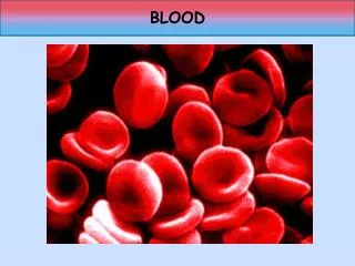
Blood • The only fluid tissue in the human body • Classified as a connective tissue • Living cells = formed elements • Non-living matrix = plasma
Physical Characteristics of Blood • Color range • Oxygen-rich blood is scarlet red • Oxygen-poor blood is dull red • pH must remain between 7.35–7.45 • Blood temperature is slightly higher than body temperature
Blood Plasma • Composed of approximately 90 percent water • Includes many dissolved substances • Nutrients • Salts (metal ions) • Respiratory gases • Hormones • Proteins • Waste products
Plasma Proteins • Albumin – regulates osmotic pressure • Clotting proteins – help to stop blood loss when a blood vessel is injured • Antibodies – help protect the body from antigens
Formed Elements • Erythrocytes = red blood cells • Leukocytes = white blood cells • Platelets = cell fragments
Erythrocytes (Red Blood Cells) • The main function is to carry oxygen • Contains hemoglobin • Anatomy of circulating erythrocytes • Biconcave disks • Essentially bags of hemoglobin • Anucleate (no nucleus) • Contain very few organelles • 4 - 6 million per mm3 of blood
Hemoglobin • Iron-containing protein • Binds strongly, but reversibly, to oxygen • Each hemoglobin molecule has four oxygen binding sites • Each erythrocyte has 250 million hemoglobin molecules
Homeostatic Imbalance • Anemia – decrease in oxygen-carrying ability of blood • May be caused by: • Low red blood cell count (RBC) • Abnormal or deficient hemoglobin • Lack of B12 or intrinsic factor • Lack of iron • Cancer • Sickle cell anemia – genetically defective hemoglobin • Polycythemia– abnormal increase in RBC • May be from bone marrow cancer or living at high altitudes • Causes increased viscosity of blood
Leukocytes (White Blood Cells) • Crucial in the body’s defense against disease • These are complete cells, with a nucleus and organelles • Able to move into and out of blood vessels (diapedesis) • Can move by ameboid motion • Can respond to chemicals released by damaged tissues
Leukocyte Levels in the Blood • Normal levels are between 4,000 and 11,000 cells per mm3 • Abnormal leukocyte levels • Leukocytosis • Above 11,000 leukocytes/ml • Generally indicates an infection • Could indicate leukemia (WBC cancer) • Leukopenia • Abnormally low leukocyte level • Commonly caused by certain drugs
Platelets • Derived from ruptured cells • Needed for the clotting process • Normal platelet count is 250,000 - 500,000/mm3
Hematopoiesis • Blood cell formation • Occurs in red bone marrow • All blood cells are derived from a common stem cell (hemocytoblast) • Hemocytoblast differentiation • Lymphoid stem cell produces lymphocytes • Myeloid stem cell produces other formed elements
Fate of Erythrocytes • Unable to divide, grow, or synthesize proteins • Wear out in 100 to 120 days • When worn out, are eliminated by phagocytes in the spleen or liver • Lost cells are replaced by division of hemocytoblasts
Control of Erythrocyte Production • Rate is controlled by a hormone (erythropoietin) • Kidneys produce most erythropoietin as a response to reduced oxygen levels in the blood • Homeostasis is maintained by negative feedback from blood oxygen levels
Hemostasis • Stoppage of blood flow • Result of a break in a blood vessel • Hemostasis involves three phases • Platelet plug formation • Vascular spasms • Coagulation
Platelet Plug Formation • Collagen fibers are exposed by a break in a blood vessel • Platelets become “sticky” and cling to fibers • Anchored platelets release chemicals to attract more platelets • Platelets pile up to form a platelet plug
Vascular Spasms • Anchored platelets release serotonin • Serotonin causes blood vessel muscles to spasm • Spasms narrow the blood vessel, decreasing blood loss
Coagulation • Injured tissues release thromboplastin • PF3 (a phospholipid) interacts with thromboplastin, blood protein clotting factors, and calcium ions to trigger a clotting cascade • Prothrombin activator converts prothrombin to thrombin (an enzyme) • Thrombin joins fibrinogen proteins into hair-like fibrin • Fibrin forms a meshwork (the basis for a clot)
Blood Clotting • Blood usually clots within 3 to 6 minutes • The clot remains as endothelium regenerates • The clot is broken down after tissue repair
Undesirable Clotting • Thrombus • A clot in an unbroken blood vessel • Can be deadly in areas like the heart • Embolus • A thrombus that breaks away and floats freely in the bloodstream • Can later clog vessels in critical areas such as the brain
Bleeding Disorders • Thrombocytopenia • Platelet deficiency • Even normal movements can cause bleeding from small blood vessels that require platelets for clotting • Hemophilia • Hereditary bleeding disorder • Normal clotting factors are missing
Blood Groups and Transfusions • Large losses of blood have serious consequences • Loss of 15 to 30 percent causes weakness • Loss of over 30 percent causes shock, which can be fatal • Transfusions are the only way to replace blood quickly • Transfused blood must be of the same blood group
Human Blood Groups • Blood contains genetically determined proteins • A foreign protein (antigen) may be attacked by the immune system • Blood is “typed” by using antibodies that will cause blood with certain proteins to clump (agglutination)
Human Blood Groups • There are over 30 common red blood cell antigens • The most vigorous transfusion reactions are caused by ABO and Rh blood group antigens
ABO Blood Groups • Based on the presence or absence of two antigens • Type A (only A antigen) • Type B (only B antigen) • Type AB (both A and B antigen) • Type O (neither A or B)
Rh Blood Groups • Named because of the presence or absence of one of eight Rh antigens (agglutinogen D) • Most Americans are Rh+ • Problems can occur in mixing Rh+ blood into a body with Rh– blood
Rh Dangers During Pregnancy • Danger is only when the mother is Rh– and the father is Rh+, and the child inherits the Rh+ factor • The mismatch of an Rh– mother carrying an Rh+ baby can cause problems for the unborn child • The first pregnancy usually proceeds without problems • The immune system is sensitized after the first pregnancy • In a second pregnancy, the mother’s immune system produces antibodies to attack the Rh+ blood (hemolytic disease of the newborn)
Developmental Aspects of Blood • Sites of blood cell formation • The fetal liver and spleen are early sites of blood cell formation • Bone marrow takes over hematopoiesis by the seventh month • Fetal hemoglobin differs from hemoglobin produced after birth
The Cardiovascular System • A closed system of the heart and blood vessels • The heart pumps blood • Blood vessels allow blood to circulate to all parts of the body • The function of the cardiovascular system is to deliver oxygen and nutrients and to remove carbon dioxide and other waste products
The Heart • Location • Thorax between the lungs • Pointed apex directed toward left hip • About the size of your fist
The Heart: Coverings • Pericardium – a double serous membrane • Visceral pericardium • Next to heart • Parietal pericardium • Outside layer • Serous fluid fills the space between the layers of pericardium • Pericarditis – inflammation of pericardium, which causes a decrease in serous fluid
The Heart: Heart Wall • Three layers • Epicardium • Outside layer • This layer is the parietal pericardium • Connective tissue layer • Myocardium • Middle layer • Mostly cardiac muscle • Endocardium • Inner layer • Endothelium
The Heart: Chambers • Right and left side act as separate pumps • Four chambers • Atria • Receiving chambers • Right atrium • Left atrium • Ventricles • Discharging chambers • Right ventricle • Left ventricle
The Heart: Valves • Allow blood to flow in only one direction • Four valves • Atrioventricular valves – between atria and ventricles • Bicuspid valve or mitral (left) • Tricuspid valve (right) • Semilunar valves between ventricle and artery • Pulmonary semilunarvalve • Aortic semilunar valve
The Heart: Valves • Valves open as blood is pumped through • Held in place by chordae tendineae (“heart strings”) • Close to prevent backflow
The Heart: Associated Great Vessels • Aorta • Leaves left ventricle • Pulmonary arteries • Leave right ventricle • Vena cava • Enters right atrium • Pulmonary veins (four) • Enter left atrium
Coronary Circulation • Blood in the heart chambers does not nourish the myocardium • The heart has its own nourishing circulatory system • Coronary arteries • Cardiac veins • Blood empties into the right atrium via the coronary sinus
Homeostatic Imbalance • Valvularstenosis– valve flaps become stiff, often from endocarditis (bacterial infection of endocardium) • Faulty valves may be replaced by synthetic or pig valves • Angina pectoris – chest pain as a result of the myocardium being deprived of oxygen • Myocardial infarction (heart attack) – prolonged angina that kills heart cells


