Case Histories
Case Histories. Case 1: Positive Newborn Metabolic Screen. History : NMS test result shows elevated Phenylalanine (0.75 umole/l; normal <0.125) Term pregnancy Normal P/L/D BWt 3.1 kg, Normal neonatal course Questions:


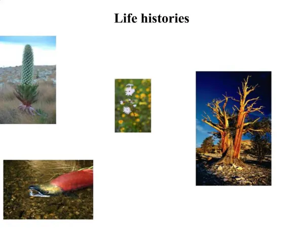
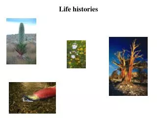
Case Histories
E N D
Presentation Transcript
Case 1: Positive Newborn Metabolic Screen History: • NMS test result shows elevated Phenylalanine (0.75 umole/l; normal <0.125) • Term pregnancy • Normal P/L/D • BWt 3.1 kg, • Normal neonatal course Questions: • Describe briefly what your initial counselling to parents would be. • What investigations would you under take to confirm diagnosis?
Results Plasma PHE=1.2umole/l; tyrosine = 0.05 umole/l Urine organic acids increased PPA,PLA,PAA Questions What other tests need to be done to be sure this baby needs diet treatment? 2. What is the basis of the diet treatment? Results of investigations
Metabolism of Phenylalanine Dietary Protein Body Protein PHE BH4 (PAH) PPA qBH2 6-pyruvoyl-BH4 Tyrosine PAA PLA GTP Phenylactetyl- glutamine
PHE levels in the Newborn with PKU Screening Possible PHE NORMAL RANGE 1 2 3 4 Days of Age
Untreated Phenylketonuria Signs / Symptoms • mental retardation • hypopigmentation • eczema-like rash • autistic-like behavior • autosomal recessive • high blood phenylalanine levels
PKU: Diagnostic work-up • Confirm that PHE level is elevated • Rule out biopterin deficiency disorders • Urine pterin levels • Dihydrobiopterin reductase activity • Biopterin load test (optional) • If present start DOPA/carbiDOPA/5HTP • If BH4 disorder not diagnsosed & PHE above 0.4 mM/l, start low PHE diet
Nutritional Treatment of PKU • Diet has two components: • Must meet all nutritional needs + limit intake of restricted nutrients to amts sufficient for growth Medical Formula Contains all nutrients except those being restricted Natural Foods Contains some normal nutrients and all those being restricted +
Case 2: Hepatomegaly with Abnormal Liver Pathology History: • The pathologist in your hospital calls to discuss an abnormal liver biopsy result • 8 month old boy with (R) abdominal mass extending down into the iliac fossa • Seen by Oncology re: ? Tumor; taken to OR for open biopsy • Question • What do you see in the following biopsy?
Questions • What types of disorders might cause this appearance? • What further historical information may be of help? • What further studies should you request from the pathologist?
Questions • What types of disorders might cause this appearance? • Glycogen storage disorders (types 1a & 1b, 3, 6 lysosmal storage disorders (Gaucher, Niemann-Pick, MPS, oligosaccharidoses, tyrosinemia, B-oxidation disorders (MCAD, LCHAD, VLCAD) • What further historical information may be of help? • Symptoms of hypoglycemia (relationship to fasting including timing) • Mother indicates baby can only go about 2-4 hours without a “bottle” • What further studies should you request from the pathologist? • PAS staining +/- pretreatment with diastase • Electon microscopy
Diagnostic testing • Fasting challenge +/- feeding challenges • Enzyme assays • Need fresh liver • Need to choose specific enzymes to target based on history • Molecular testing • Now have bank of mutations but expensive
GSD-II ( lysosomal) GSD-IV GSD-V,GSD-VI,GSD-IX GSD-0 GSD-III GSD-1a&b GSD-VII GSD-X,GSD-XII,GSD-XIII GSD-XI (LDH) LIVER MUSCLE
Clinical features early onset hypoglycemia lactic acidosis hepatomegaly Fanconi syndrome hyperuricemia hyperlipidemia Diagnosis controlled fast (test BS & LA) enzyme (liver biopsy) DNA testing Therapy provide 5 - 10 mg glucose/kg/min continuous .nocturnal infusion of CHO as polycose or formula frequent meals during days corn starch days &/or nights don’t over treat with CHO Neutropenia in Type IB prophylactic antibiotics GCSF Emergency protocols for illness, surgery etc. GSD IA &IB
Case 3: 18 month boy with hepatomegaly and obtundation History: • stuporous on admission • found pale & sweaty and unarousable by parents • Had been ill for about 18 hours with no significant intake • Seizure in ambulance Initial studies: • Blood sugar = 0.2 mM/l, Na+=145, K+ =3.5, Cl-=104, TCO2 = 10 • Urinalysis = Normal • All other testing including lactate, NH4 & LFE’s normal
Key observations • Severe hypoglycemia with hepatomegaly and no ketonuria on setting of history of prolonged fasting • Needs urgent treatment of hypoglycemia • Route? • How much glucose? • ? Significance of no ketones in urine • ?diagnostic testing
VLCAD,MCAD, SCAD Trifunctional protein
Diagnostic Investigations • Plasma acylcarnitnes suggest Medium Chain Dehydrogenase deficiency (MCAD) • Plasma free carnitine levels low while acylcarnitines high • 14C- palmitic acid oxidation in leucocutes quite reduced • Molecular diagnosis indicates homozygosity for the common caucasian mutation.
Phases of Glucose Homeostasis 1.Glucose absorptive phase: 3 - 4 hrs after glucose ingestion (high insulin) 2.Post absorptive/early starvation: 3-12 hrs glucose (from hepatic glycogen) to brain, RBC, renal medulla 3. Early / Intermediate Starvation: 14+ hrs gluconeogenesis & (later) lipolysis
Treatment • Avoid fasting • L-carnitine if free carnitine low • Emergency protocol & letter • Sick day management • Admission to ER/hospital to maintain blood glucose with IV infusion to prevent excessive lipolysis the would overload the B-oxidation pathway
5 d.o. male Well for 72 hrs then became lethargic, fed poorly, began vomiting & developed alternating flaccidity & opisthotonic posturing. Became comatose Developed hyperpnea and respiratoy alkalosis progressing to respiratory failure O/E: hepatomegaly, hypothermia Test Results Normal: CBC, ‘lytes’, bld glucose, lactic acid, urinalysis not acidotic not ketotic not hypoglycemic NH3 (350 umole/l) ? Differential Diagnosis ? Further testing Case 4: Neonatal Presentation
5 d.o. male Well for 72 hrs then became lethargic, fed poorly, began vomiting & developed alternating flaccidity & opisthotonic posturing. Became comatose Developed hyperpnea and respiratoy alkalosis progressing to respiratory failure O/E: hepatomegaly, hypothermia Test Results Normal: urine amino acids & organic acids Low: urea, arginine, ornithine, High: citrulline (1.21 mM) glutamine (1.4 mM) asparagine ? Probable Diagnosis Case 4: Investigation Results
Detoxification of NH3 by Urea Cycle Benzoate Dietary Protein Gut Bacteria Endogenous Protein Catab Buphenyl NH4 PAA NH4 + CO2 GLN Carbamoyl Phosphate GluAPhAcGluNH2 HippuricAcid Ornithine Citrulline MITOCHONDRION Aspartate Urea Arginine Argininosuccinic Acid Fumarate (CPS) (OTC) (Arginase) (ASAS) (ASAL)
Approach To Differentiation of Urea Cycle Disorders Causing Neonatal Hyperammonemia Hyperammonemia Before 24 hrsAfter 24 hrs Metabolic acidosis Preterm Fullterm YesNo plasma citrulline THANPDH PAA abs/tr>1000uMGAIIMMA 100-300uM etc.etc. Ur. Orotate uOA low high ASA CPS OTCCitrullinemia
Acute Mgmt (based on NH3 level) NPO Dialysis ( prefer. Hemodialysis) IV: CHO (6–8 mg Glc/kg/min) Lipid (3 gm / kg) Alternate Pathway Therapy Oral (Phenylbutyrate + L-Arg) IV (Phenylacetate + benzoate + L-arginine Chronic Mgmt Low protein diet –1.0 to 1.5 gm/kg/d -Cyclinex (ess. AA’s) (up to 50 % of prot) Phenylbutyrate (Buphenyl) (450-650mg/kg/d) Arg / ornith / citrulline Regular monitoring Approaches to Therapy of Urea Cycle Disorders
Case 5: 5 year old girl with hearing loss and macrocephaly • Relatively normal global development otherwise • Family history negative & parents unrelated • S/S hepatomegaly, mild contractures-hands, knees & elbows
Case History: HM • You were asked to see this girl for assessment regarding macrocephaly and hearing loss • Initial investigations showed an abnormal urine metabolic screen that positive for both CTAB & Toluidine blue • All other initial metabolic studies normal • What would you do next?
“Coursened Features”Laboratory Screening Paradigm MPS Storage Disease Uronic acid Yes MPS by TLC Enzyme Assays Course facies MPS in urine MLS II or III Total Hex Oligo screen No Oligosaccharidoses Enzyme Assays
Uronic acid GAG electrophoresis How do you measure mucopolysaccharide excretion? What are the useful GAGS in diagnosing a particular MPS disorder? How is this testing done?
Uronic acid GAG electrophoresis How do you measure mucopolysaccharide excretion? A B Acid Hydrolysis to free monosaccharide subunits + Colorimetric detection of the uronic acid (A or B)
Urine MPS excretion Uronic acid by acid hydrolysis /carbazole Uronic acid is 52 ug/ml (Normal < 16.7) Other methods GAG electrophoresis ( HM spec) Additional testing
Diagnosis & Management • Mucopolysaccharidosis Type III • San Filippo syndrome • Four different enzyme deficiencies all leading to inadequate breakdown of heparin sulfate • She had deficiency of heparin-N-sulfatase (MPS-IIIA) • No definitive treatment at present • extensive social/psychological/educational support • appropriate for child with neurodegenerative disorder
History • The mother of a six year old boy tells you that her son has had three episodes of abdominal pain without any flu-like symptoms or other systemic problems. • During the last episode, blood but no bacteria or “pus” cells were seen in his urine. The urine did not smell, look cloudy or have an unusual color. • Her husbands had had similar episodes as a child but these had stopped once he followed the advice of a pediatrician to ”drink lots of water” at nights.
Your initial investigations showed that this boy had a normal physical exam, normal CBC. ‘lytes, urea & creatinine and aside from microscopic hematuria, a normal microscopic & chemical urinalysis • What do you think is causing his abdominal pain & microscopic hematuria? • What genetic / metabolic disorders should you consider in your differential diagnosis list? • What specific testing would you suggest to investigate these possibilities?
Differential Diagnostic List • Cystinuria (basic aminoaciduria) • Partial HGPRTase deficiency (uricosuria) • GSD I (uricosuria) • Primary hyperoxaluria (oxalic aciduria +/- glycolic or glyceric acid) • Idiop. Hypercalciuria (calcium oxalate or urate) • Hyperparathyroidism (calcium oxalate / urate) • Adenosine phosporibosyl transferase deficiency (dihydroxyadenosinuria)
Results of Special Testing • Unable to isolate characteristic stones in random urine specimen • Urine oxalic acid, calcium, uric acid / creatinine ratio all normal • urine oxypurine profile normal • cystine, lysine, ornithine & arginine but no other amino acids elevated in urine • Plasma amino acid profile normal • What is your Diagnosis?
disorder of basic amino acid transport involving renal tubule & GI mucosa urolithiasis:poor solubility of cystine when: urine concentrated (cystine> 1200 uM/l) urine acidic autosomal recessive 1: 7000 Types I, II & III (newer classification refers to type I as classical & others as non-classical) Heterozygotes (type II) may excrete Cyst. Lys & Orn +/- arg but in reduced amts Renal immaturity (< 1 yr) may cause “apparent” cystinuria in Type III carrier Cystinuria
Dibasic Amino Aciduria • Normal:Cystine filtered at glomerulus but over 98 % reabsorbed in proximal renal tubule • Common renal tubular reuptake mechanism for dibasic amino acids (cystine,lysine, ornithine, arginine). All increased in urine but not blood if transporter is deficient • Nephrolithiasis: onset by 10 yrs (25-30 %) to 20 yrs (50-60 %) • Cystine precipitates out in urine / renal filtrate at concentrations above 1200 uM/l • stones: hexagonal, golden-brown, grain size to staghorn size • account for 1-3 % of all stones in adults (kids ?) • Subtypesbased on amt excretion of cys/lys/orn but not arg in heterozygotes. Type III has similar defect in mucosal uptake
Locus 1: 2p16.3-p21 Type I cystinuria rBAT protein SLC3A1 gene 40+ mutations Transmembrane protein Locus 2: 19q13.1 Non-Type 1 cystinuria Bo, +AT protein SLC7A9 gene 30 + mutations Complexes with rBAT protein to form dibasic amino acid transporter Molecular Genetics
Phenotype/Genotype Correlation • Cystinuria I/I: • two fully recessive SLC3A1 mutations • no gut absorption • kidney - high risk for nephrolithiasis • Cystinuria III/III • two incompletely recessive SLC7A9 mutations • stones in adults • some gut absorption • Mixed types: I/III(A), I/III(B), II/II,
Treatment of Cystinuria • dilution of urine: • drink up to 4.0 liters fluid / day (1.75 - 2.0 l/m2/24 hr) • important to drink during the night time (water at bedside) • monitor urine cystine concentrations morning & evening • “alkalinization” of urine: NaHCO 3 ( 1.5 - 2.0 mEq/kg/24 hr) • Medication: D-penicillamine, Thiola R (tiopronin) • Surgical: lithotripsy etc.