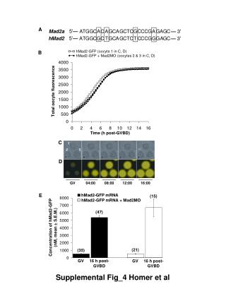
Fluorescence Study of hMad2-GFP in Oocytes with Mad2 Morpholino Treatment
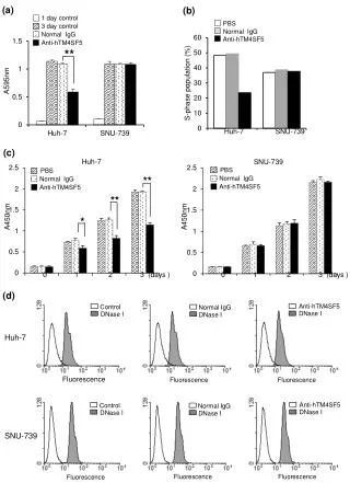
This study investigates the effects of hMad2-GFP and Mad2 Morpholino in oocytes, focusing on total fluorescence over time post-GVBD. Analyzed samples include hMad2-GFP mRNA conditions and concentrations, with fluorescence measured at various intervals (0, 4, 8, 16 hours). Results are supplemented with graphical data detailing fluorescence intensity and corresponding statistical analysis. This research contributes to understanding the role of hMad2 in oocyte maturation when influenced by genetic perturbations.

Fluorescence Study of hMad2-GFP in Oocytes with Mad2 Morpholino Treatment
E N D
Presentation Transcript
hMad2-GFP (oocyte 1 in C, D) hMad2-GFP + Mad2MO (oocytes 2 & 3 in C, D) C 1 2 3 D 04:00 08:00 16:00 GV 12:00 5'— C A T G G C A C A G C A G C T C G C C G A G A G C — 3' Mad2a 5'— A T G G C G C T G C A G C T C T C C C G G G A G C — 3' hMad2 A B Total oocyte fluorescence Time (h post-GVBD) E hMad2-GFP mRNA (15) hMad2-GFP mRNA + Mad2MO (47) Concentration of hMad2-GFP (nM, mean S.E.M.) (21) (35) GV 16 h post- GVBD GV 16 h post- GVBD Supplemental Fig_4 Homer et al