Download
1 / 17
240 likes | 704 Vues
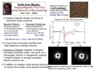
Resonant magnetic x-ray scattering and Summary. Resonant scattering Why do it? What is it? How is it done? Example(s) The Real World …. CaFe 2 As 2. Solving magnetic structures. Determine the magnetic wavevector (what is the “magnetic unit cell”)

E N D
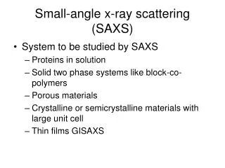
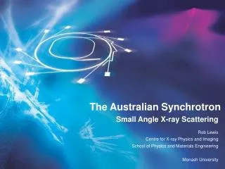
Resonant magnetic x-ray scattering and Summary • Resonant scattering • Why do it? • What is it? • How is it done? • Example(s) • The Real World …. CaFe2As2
Solving magnetic structures Determine the magnetic wavevector (what is the “magnetic unit cell”) Use the angular dependence of resonant and nonresonant scattering cross-sections to determine magnetic moment directions. Scattering amplitudes → magnitude of the ordered magnetic moment.
Long-range order Bragg peaks t = 2p/2d 1 t t 10-6 2d QBragg=2p/d I d 2p/d Q
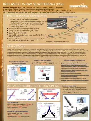
Why Bother? • Many of the technologically important RE compounds contain neutron opaque elements. • Superior reciprocal space (Q) resolution allows more detailed study … reinvestigation of “solved” structures. • Can be used for investigations of submillimeter-sized single crystals. • Resonant magnetic scattering occurs at well-defined energies specific to elements of interest -- probe local magnetism. • Studies of magnetic surfaces and interfaces.
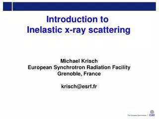
L3 - edge EF outgoing photon Incoming photon P3/2 P1/2 X-ray Resonant Magnetic Scattering (XRMS) • (L2, L3)-edge for rare-earths (6-10KeV) • Electric multipole transition • (dipole : 2p – 5d, quadrupole 2p – 4f) • Dipole transition is dominant • 4f : magnetic properties • 5d : exchange splitting by 4f
analyzer sample Non-resonant: ds/dW [S2sinQ]2(outgoing s- pol.) [2sin2Q cosQ{(L1+S1) + S3sinQ}]2(outgoing p- pol.) Resonant (E1): ds/dW 0(outgoing s- pol.) (-M1cosQ + M3sinQ)2(outgoing p- pol.) (k′· M)2 Resonant (E2): Much more complicated, but can probe M1, M2, M3 ^ ^ ^
We can do this by plotting the q-dependenceof integrated intensities (a la neutrons) Angular dependence of the scattering at (0 0 L ± t) of GdCo2Ge2 measured by resonant and nonresonant diffraction Gd L3 edge Alternatively…
Q k' k θf θi X-ray resonant magnetic scattering azimuth scans Gd5Ge4 Intensity µ |k'• M|2 I (M•k’)2
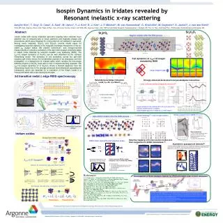
Crystal structure GdNi2Ge2 – An Example Magnetization measurement TN Tt Gd Ni Ge S.L. Bud’ko, Z. Islam, T.A. Wiener, I.R. Fisher, A.H. Lancerda, P.C. Canfield Journal of Magnetic Materials 205, 53 (1999)
1st harmonic (magnetic) 2nd harmonic (charge) 3rd harmonic (magnetic)
So, what do you learn from diffraction? • From peak positions • Lattice parameters and how they change with environmental conditions (e.g. temperature and pressure) • From peak widths • Crystal quality (e.g. mosaic) • Presence of strain (e.g. longitudinal widths) • From integrated intensities • Contents of the unit cell • Positions of atoms within the unit cell; magnetic ordering • Thermal parameters (thermal disorder)
Peak Widths – strain, crystallite size and mosaic Powder after grinding Single crystal mosaic