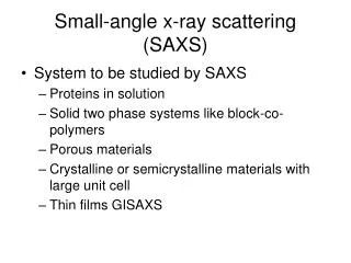
SAXS Small Angle X-ray Scattering
SAXS Small Angle X-ray Scattering. Talia Zemach (2002) Raanan Kalifa (2003) Rami Shnaiderman (2005) Materials Engineering, BGU Lecturer: Prof. Jacob Zabicky. Introduction—the scale of things. Scattering from X-rays Neutrons Electrons Light


SAXS Small Angle X-ray Scattering
E N D
Presentation Transcript
SAXSSmall Angle X-ray Scattering Talia Zemach (2002) Raanan Kalifa (2003) Rami Shnaiderman (2005) Materials Engineering, BGU Lecturer: Prof. Jacob Zabicky
Introduction—the scale of things Scattering from • X-rays • Neutrons • Electrons • Light is used in many disciplines to study a vast range of materials.
Introduction—light scatterimg Electromagnetic radiation can be used to obtain information about materials of dimensions of the same order as the wavelength of the radiation passing though the material. For example, an ordinary glass of milk vividly illustrates this principle. In normal visible light the milk appears as a continuous, ‘milky’ white fluid. However, place the same glass of milk in ultraviolet “black” light and it appears as an emulsion of particles because the wavelength of the UV light is similar to the dimensions of the butterfat that is dispersed as tiny drops in the serum.
Introduction—X-rays scatterimg Around the turn of the 20th century, Röntgen discovered radiation with wavelength much shorter than that of visible light (less than 1 nm) – the X-rays. Soon after that von Laue and his associates discovered that crystals scatter X-rays in distinct patterns (diffraction). It was soon recognized that these patterns give direct insight into the structure of the materials that caused the scattering, allowing us to determine the morphology and size of the particles (e.g., molecules) assembling the mater. Scattering experiments usually consist of irradiating a sample with highly coherent X-ray radiation and measuring the resulting scattering pattern.
The reciprocity law The reciprocity law: The larger the diffraction angle the smaller the length scale probed and viceversa. Wide angle X-ray scattering (WAXS) is used to determine crystal structure on the atomic length scale while small-angle X-ray scattering (SAXS) or small-angle neutron scattering (SANS) are used to explore microstructure on the colloidal length scale .
The reciprocity law The larger the diffraction angle the smaller the length scale probed and vice versa. The difference between small angle scattering (SAXS) and wide angle scattering (WAXS) is in the scale of the sample and consequently the size of the angle. • – wavelength of X-ray d – distance between relevant parts of the analyzed sample • – half the angle between the diffracted and transmitted beams For example, d = 250 Å λ = 1.5 Å (fixed) corresponds to = 0.17° = = 0.030 rad
What is SAXS? Small angle X-ray scattering (SAXS) is a well-established chatacterization tool that has been around for about 70 years. It is "special" in terms of the distinction between SAXS and regular wide-angle X-ray scattering by virtue of the location of the scattering of interest. This is typically at small angles in the vicinity of the primary beam and extending to less than 2 degrees for standard wavelengths. The scattering features at these angles correspond to structures ranging in size from tens to thousands of angstroms. • Closer inspection shows that the X-ray is diffracted due to regularly spaced scattering objects, e.g. atoms in a solid matter, that cause differences in electron density. For diffraction to occur the spacing between the objects must be equal to an integral multiple of the wavelength of the probing radiation, making the X-rays perfect for investigation of nano-structures.
Why not light scattering? • Frequently we need to look at bulk materials which are opaque to visible light. • The wavelength of light is much larger than that of X-rays (or neutrons), which makes light scattering adequate for much larger structures (like phases in blends of elastomers). Why not electron microscopy ? • The need to produce very thin slices for electron microscopy can destroy the very thing we want to observe. Why SAXS? • While SANS is usually a possibility, it certainly is less straightforward than SAXS. • There are cases where SAXS is either the best or the only source of information that is needed on some materials.
SAXS experiments • The electrons in the sample scatter off X-rays. • Being X-rays of high energy small scattering angles are possible. • A long instrumental setup is needed to measure the small angles. • Scattered and transmitted X-rays are detected on an area detector, for example, a charge-coupled device (CCD), and the display is sent to a computer for processing. • The scattering angle is determined from the distance of the pixel to the center of the display. • The scatering angle allows estimation of the radius of giration (Rg) and the particle shape.
The result of a SAXS experiment is essentially the intensity of the Fourier transform of the electron density and must be interpreted in order to determine morphology, size, structure, physical properties (volume, area, weight ). • Direct methods yield information based on interpretation of the clean (background corrected) data with no further manipulation. However, all these parameters are based on well-defined assumptions such as uniform density within the so-called particle, uniform density in the background, sharp interfaces between the two, etc. • When these assumptions do not apply one can carry out a Fourier transform of the data to get real space information. However, not as in electron microscopy, a fundamental problem with any scattering experiment is that two different morphologies can, in theory, give identical scattering patterns. Generally, one cannot reconstruct the exact microstructure uniquely from a SAXS pattern because in a scattering experiment only the scattered radiation intensity can be measured and all phase information is lost .
The scattering intensity is proportional to the number of scattering elements in the irradiated volume Np(1/q). Then,in small angle scattering we can consider a generalized rule that describes the behavior of scattered intensity as a function of Bragg’s size “d” or ‘r” that is observed at a given scattering angle 2θ , where r =1/q. • When these assumptions do not apply one can carry out a Fourier transform of the data to get real space information (such as obtainable by electron microscopy).
A scattering experiment The detected patterns may be isotropic, oriented, bimodal, etc.
SAXS processed results It is helpful to recognize some of the more typical SAXS patterns of isotropic systems, i.e. whether macroscopic orientation exists or not in the scattering volume.
SAXS result on the CCD and in q space A bright ring means there is a periodic structure in that object with the corresponding repeat spacing. Intense scattering over a range of angles means the structure is ordered on that length scale but not periodic.
SAXS Data Analysis-Direct methods. • Guinier region: • At very small angles, the shape of the scattering can be used to give us an idea of the radius of gyration of any distinct structures that are on this range of length scale. • At higher angles, if we had a system of relatively uniform particles, dilute enough for mutual interactions, we might be able to see broad peaks that would also give us information on the shape of the particles. Dmin = 2π/qmin q = 2πsin/
SAXS Data Analysis. • Porodregion: At higher angles, the shape of the curve gives information on the surface-to-volume ratio of the scattering objects. This can also be used to obtain information on the dimensions of the scattering particles. • INVARIANT : The area under the curve is a measure of the amount of scattering material seen by the X-ray beam. Changes in the invariant are useful to follow crystallization in polymeric materials. q = 2πsinθ/λ D max = 2π/qmax Do = 2π/qo
r Examples of data analysis-direct methods R=10 r I q-Df R, Df =2 L. Barre, IFP France
r Examples of data analysis-direct methods R=10 r I q-Df R, Df =2 L. Barre, IFP France
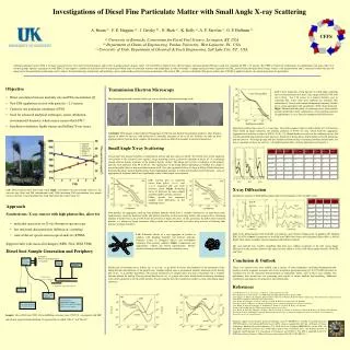
Applications Many applications of the SAXS technique are found in : • Structural biology (proteins, muscles, lipid vesicles). • Polymer science. • Colloidal chemistry. • Materials science (crystalline alloys, amorphous materials, single crystals, porous materials, liquid crystals). • Chemical analysis.
Applications • For monodisperse particle systems • Radius of gyration of particle, of rod or platelet, i.e. size parameter • Particle shape in solutionミ comparison of modelled vs. experimental scattering curve • Radial electron density distribution, e.g. of micelles • Molecular weight of particle, of unit length (rods), or unit area (plates) • For concentrated solutions • Solution structure function • For polymers • in solution: persistence length, degree of coiling • in bulk: domain structure (crystalline vs. amorphous), lattice structure • For liquid crystals ミ low-dimensional systems • Lattice symmetry and dimensions • Degree and nature of long-range disorder • For powders • Specific inner surface / surface-to volume ratio of inhomogeneities
Applications A SAXS pattern of a surfactant templated mesoporous silicate
Applications Latex particles of uniform size in solution can be described as a dense core surrounded by a hairy halo of loose tails.
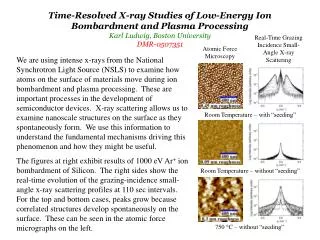
Applications In the presence of Ca(II), gelsolin undergoes a structural change that allows recognition and binding to actin.
Applications Diffraction pattern of a highly aligned lipid (DMPC) in the Pbォ phase at constant grazing angle at 20 °C. Letter A shows the in-plane reflections due to the ripple structure (distance lambda), letter B gives the off-plane reflections due to the lamellar repeat distance d (see lower insert). C denotes the specular reflectivity peak.
To Sum it up… • SAXS allows studying nano-structured materials (1-100nm). on their typical length scale. • The problem of small-angle analysis consists of deducing the structural features, such as size, shape or mass, from the interference pattern.
To Sum it up - Main advantages. • Good statistics: the volume of the material which is sampled is in the range from 105 to 107µm3, depending on the material, wavelength of the X-rays and focusing of the beam. Thus a very large number of particles are averaged in the measured data. • Quantitative measurements: from a SAXS spectrum, an average size of the particles can be obtained in a straightforward and automated manner. The volume fraction of the objects can be obtained too, provided that their composition is well known. • In-situ measurements: the sample size and the measuring times enable easy real time studies during heating or deformation, and detailed information can be obtained on precipitation kinetics or phase changes during straining. • Nondestructive for polymers.
A recent development The use of synchrotron radiation sources in SAXS studies increases the resolution of the experiments and decreases the acquisition times, leading the way for new types of experiments, notably those involving fast in situ measurements (with events of the order of a few seconds), in temperature or deformation, yielding time-resolved measurements as well as space-resolved studies of heterogeneous samples.
References • http://physics.queensu.ca/~marsha/SAXS.html • http://coecs.ou.edu/Brian.P.Grady/saxs.html • http://www-ssrl.slac.stanford.edu/~saxs/ • http://www.molmet.com • http://www.jjxray.dk/sxupg.html • http://www.hecus.at/image/i_c_10_02.gif • http://scattering.tripod.com/ • Encyclopedia of Materials, Microscopic, spectroscopic and physical techniques • Michette/Buckly, X-Ray Science and Technology, 1993. • A small angle X-ray Apparatus for studying biological macromolecules solution, J. Appl. Cryst., (1998) 31, 533-543. • Kirk-Othmer’s Encyclopedia of Industrial Chemistry, Structure analysis by diffraction.