Download
1 / 20
220 likes | 288 Vues
Explore the world of small angle scattering, from crystal structures to gas transport properties, using examples such as block copolymers and SOFC layers. Learn about critical dimension metrology in nanoscale structures and wood cell walls.

E N D
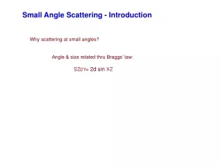
Small Angle Scattering - Introduction Why scattering at small angles? Angle & size related thru Braggs’ law: = 2d sin
Small Angle Scattering - Introduction Why scattering at small angles? Angle & size related thru Braggs’ law: = 2d sin Suppose CuK x-rays (= 1.5418 Å) calculate 2 for various ds
Small Angle Scattering - Introduction Why scattering at small angles? Angle & size related thru Braggs’ law: = 2d sin Suppose CuK x-rays (= 1.5418 Å) calculate 2 for various ds d 2 q 10 Å (0.001 micron) 8.84° 0.628 Å-1 50 Å 1.77° 100 Å (0.01 micron) 0.88° 0.0628 Å-1 300 Å 0.29° 600 Å 0.15° 1000 Å (0.1 micron) 0.09° 0.00628 Å-1 10,000 Å (1 micron) 0.009° 0.000628 Å-1
Small Angle Scattering - Introduction d 2 q = (4π/) sin 2π/d 10 Å (0.001 micron) 8.84° 0.628 Å-1 50 Å 1.77° 100 Å (0.01 micron) 0.88° 0.0628 Å-1 300 Å 0.29° 600 Å 0.15° 1000 Å (0.1 micron) 0.09° 0.00628 Å-1 10,000 Å (1 micron) 0.009° 0.000628 Å-1 High-angle x-ray scattering (usually ~2°-160°2) --> atomic scale structure
Small Angle Scattering - Introduction d 2 q = (4π/) sin 2π/d 10 Å (0.001 micron) 8.84° 0.628 Å-1 50 Å 1.77° 100 Å (0.01 micron) 0.88° 0.0628 Å-1 300 Å 0.29° 600 Å 0.15° 1000 Å (0.1 micron) 0.09° 0.00628 Å-1 10,000 Å (1 micron) 0.009° 0.000628 Å-1 High-angle x-ray scattering (usually ~2°-160°2) --> atomic scale structure Small-angle scattering --> structure of BIG things
Small Angle Scattering - examples WAXS and SAXS study of (m)TMXDI-PDMS siloxane-urethaneureas
Small Angle Scattering - examples WAXS and SAXS study of (m)TMXDI-PDMS siloxane-urethaneureas Hard segments --> regions with crystal-like order Soft segments --> amorphous siloxane chains
Small Angle Scattering - examples WAXS and SAXS study of (m)TMXDI-PDMS siloxane-urethaneureas Saxs scattering curves for various NCO/OH ratios (a = 1.5/1; e = 4.5/1): Size of the hard segment “crystalline” regions changes 0.9°
Small Angle Scattering - examples Microstructure orientation and nanoporous gas transport in semicrystalline block copolymer membranes Polymer sheets of semicrystalline ethylene (E)/ethylene–propylene (EP) diblock E/EP and triblock E/EP/E copolymersmade by channel die proessing
Small Angle Scattering - examples Microstructure orientation and nanoporous gas transport in semicrystalline block copolymer membranes Polymer sheets of semicrystalline ethylene (E)/ethylene–propylene (EP) diblock E/EP and triblock E/EP/E copolymersmade by channel die processing Sheets are stacked into several types of blocks w/ different gas transport props.
Small Angle Scattering - examples Microstructure orientation and nanoporous gas transport in semicrystalline block copolymer membranes Polymer sheets of semicrystalline ethylene (E)/ethylene–propylene (EP) diblock E/EP and triblock E/EP/E copolymersmade by channel die processing FD CD 145 Å 436 Å saxs image for perpendicular texture type
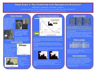
Small Angle Scattering - examples Nanometer to Micrometer Void Microstructure Characterization of SOFC Layers and Interfaces by Small Angle Scattering (SAXS) and Computed X-ray Microtomography(XMT)
Small Angle Scattering - examples Critical Dimension Metrology of Nanoscale Structures with Small Angle X-ray Scattering NIST developing transmission saxs method capable of angstrom level precision in critical dimension evaluation over (50 x 50) mm arrays of nanoscale periodic structures
Small Angle Scattering - examples Critical Dimension Metrology of Nanoscale Structures with Small Angle X-ray Scattering SEM image of a photoresist grating on a silicon wafer & resulting 2-D SAXS image
Small Angle Scattering - examples Critical Dimension Metrology of Nanoscale Structures with Small Angle X-ray Scattering SEM image of a photoresist grating on a silicon wafer & resulting 2-D SAXS image Streaks tell about deviations from ideal grating & defects such as long wavelength line edge roughness
Small Angle Scattering - examples Critical Dimension Metrology of Nanoscale Structures with Small Angle X-ray Scattering blue rectangles represent etched regions in a film (b) resulting SAXS detector image
Small Angle Scattering - examples The measurement of the micro-fibril angle in soft-wood Wood cell wall consists of bundles of a crystalline arrangement of cellulose chains (microfibrils)
MFA Small Angle Scattering - examples The measurement of the micro-fibril angle in soft-wood Wood cell wall consists of bundles of a crystalline arrangement of cellulose chains (microfibrils) Microfibrils align quite parallel in a spiral around cell wall, with spiral axis along long cell direction X
Small Angle Scattering - examples The measurement of the micro-fibril angle in soft-wood Typical saxs patterns from Norway spruce - mean MFA of 20° - longitudinal cell axis vertical. (c) pattern recorded at α= 0°. (d) pattern recorded at α= 45°