Download
1 / 23
250 likes | 624 Vues
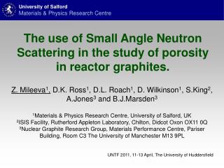
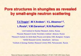
Pore structures in shungites as revealed by small-angle neutron scattering. T.V.Tropin 1 , M.V.Avdeev 1 , V.L.Aksenov 1,2 , L.Rosta 2 , V.M.Garamus 3 , N.N.Rozhkova 4 1 Joint Institute for Nuclear Research, Dubna, Russia 2 Russian Research Center “Kurchatov Institute”, Moscow, Russia

E N D
Pore structures in shungites as revealedby small-angle neutron scattering T.V.Tropin1, M.V.Avdeev1, V.L.Aksenov1,2, L.Rosta2, V.M.Garamus3, N.N.Rozhkova4 1Joint Institute for Nuclear Research, Dubna, Russia 2Russian Research Center “Kurchatov Institute”, Moscow, Russia 3Research Institute for Solid State Physics and Optics HAS, Budapest, Hungary 4GKSS Research Centre, Geesthacht , Germany 5Institute of Geology Karelian Research Centre RAS, Petrozavodsk, Russia “Stress and Textures Investigation by Means of Neutron Diffraction”, STI-2011, FLNP, JINR, Dubna, Russia
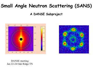
Outline • Introduction: shungite spreading, properties, applications and microstructure. • Experiments • Method: small-angle neutron scattering (SANS) • Samples description • SANS: characteristics of levels • SANS: open/close porosity study • Conclusions
Shungite spreading Shungites are carbon-rich rocks of Precambrian age widespread over Russian Karelia. There are four types of shungite different in carbon content. Buseck PR, et al. Canadian Mineralogist (1997)
Shungite deposits Shunga Maksovo
Shungite deposits Negozero
Characterization of shungites Elastic modulus 24 GPa (shungite I)Tensile strength ~110 MPa Compression strength>200 MPaDensity 1.9 g/cm3Resistance ~0.1 Ω·cm (shungite I, semiconductor) Mixture of properties Glassy carbon Coal Graphite Coke
Shungite carbon applications • low-temperature catalysts of hydrogenation • adsorbents and filters in water purification • multi-functional fillers of polymeric and inorganic binders • radiation screening construction materials • alternative material for coke and quartzite • natural source of fullerenes (?) • Buseck PR, Tsipursky SJ, Hettich R. Fullerenes from Geological Environment. • Science (1992) A general task of the study of shungites is to find out the most effective technologies of their treatment and application! …In this connection, the knowledge about the structure of this rock and its modifications is of great importance.
Shungite microstructure: HRTEM Shunga Maksovo Shungite carbon contains packed globular or ellipsoidal multi-layered graphene units (size < 10 nm) forming aggregates with chaotic (Shunga) or preferable (Maksovo) orientation. Additional study by: chemical analysis, chromatography, electron and X-ray diffraction, HRTEM, AFM, SAXS.
Research motivation • To use SANS method to reveal structural features of shungite powders at the scale of 1-100 nm complementary to previous investigations; • Compare SANS signals from shungites at different deposits and to determine characteristic parameters of various types of the structural organization of shungites; • Use the SANS contrast variation technique to separate effects of the closed and open porosity on the scattering and conclude about the pore structures of shungite.
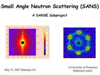
Small-angle neutron scattering size ~1-100 nm ~ 0.001-1 Instruments YuMO time-of-flight setup, IBR-2 Reactor, FLNP, JINR, Dubna Yellow Submarine steady-state setup, Budapest Neutron Center SANS-1 steady-state setup, GKSS Research Centre, Geesthacht
Contrast variation the core is matched the shell is matched
Samples Shungite carbon-rich rocks type-I, dry grinding down to granule size ~ 40 m
Small-angle neutron scattering Level I (large q-values). Surface fractal (dimension DS). Level II (small q-values). Mass fractal (dimension DM).
SANS fitting models: single level C – is the background remaining after corrections; P – characterizes the type of mass or surface organization of the scattering units; n, V, ρ – particle number density, volume and scattering length density of the scattering units; - mean scattering length density of the bulk medium; k – an empirical constant; R– radius of gyration of the particles; S – surface area of the scattering units; This model was used to fit a single level and obtain the parameters.
SANS fitting models: generalization for two levels level I level II Impact of scattering from level-II units on the scattering from level-I units. The proposed model describes the two-level structure of shungites. It allows to obtain structure parameters after the fit.
Comparison: SANS and SAXS results • The results of SANS and SAXS experiments are in a good agreement; • The fit of SANS results by the proposed models of two-level structure has been successful;
Characteristics of the levels • Level I: • n<1013 cm-3; • Volume fraction - ~4%; • Inner surface ~0.3 m2/g; • Level II: • n<1017 cm-3; • Volume fraction - <10%;
Two-level organization of shungites AFM Maksovo: HRTEM Arrow shows pore between BSU filled with non-structured fractal carbon Arrow shows large globule unit Arrows show basic structural units (BSUs) Kovalevski V.V., Prikhodko A.V., Buseck P.R., Carbon 2005; Rozhkova N.N., Golubev E.A., V.I.Siklitski, M.V.Baidakova In: Fullerenes and fullerene containing materials, Eds. Vityaz’ P.A., et al., Minsk: UP”Tehnoprint”, 2002;
Matching of open pores in shungites + D2O open pores: = D2O– C ~ 0.71010 cm-2 = – C ~ –71010 cm-2 closed pores: = – C ~ – 71010 cm-2
Matching of open pores • Comparison of the scattering curves for initial sample (Maksovo, Shunga) and the same sample after absorption of D2O during one week before experiments. • Changes in the scattering are due to matching of open porosity.
Contrast variation in shungites Pores at level I. Size > 200 nm, fraction < 10 vol. %, fully open; Open pores at level II. Size 5-10 nm, posses inner fractal structure of units less than 1 nm in size. Closed pores at level II Repeat sample elongation texture, L > 60 nm, R < 2 nm Main contribution into inner surface comes from open poresat level II: S ~ 130 m2 g-1 (SANS) S ~ 200 m2 g-1 (BET) Level I Level II
Two-level organization of shungites AFM Maksovo: HRTEM Arrow shows pore between BSU filled with non-structured fractal carbon Arrow shows large globule unit Arrows show basic structural units (BSUs) Kovalevski V.V., Prikhodko A.V., Buseck P.R., Carbon 2005; Rozhkova N.N., Golubev E.A., V.I.Siklitski, M.V.Baidakova In: Fullerenes and fullerene containing materials, Eds. Vityaz’ P.A., et al., Minsk: UP”Tehnoprint”, 2002;
Conclusions • Complex structural organization of shungites is detected in SANS, SAXS and EM experiments; • All samples from different deposits are characterized by two scattering levels corresponding to fractal structures at the scales of ~10 and > 100 nm, respectively; • Despite the structural similarity a significant difference in bulk morphology for shungites from different deposits is observed; • Open porosity determines the fractal organization of the first scattering level (size ~10 nm), while the close pores at this level are polydisperse elongated globules, which is in agreement with the present model of shungite basic structural units.