Download
1 / 34
340 likes | 382 Vues
Learn about DNA fingerprinting using restriction enzymes to cut DNA at specific sites, then visualize the fragments through gel electrophoresis for analysis. Understand the history, classes of endonucleases, naming conventions, and techniques involved in this fascinating process.

E N D
Restriction Enzyme DigestionDNA Scissors Restriction endonucleases, known as restriction enzymes or REs, are protein molecules that cut DNA at specific locations, called restriction sites, in their nucleotide sequence.
History • Restricting, or cutting of DNA, was first observed in 1950s when certain strains of bacteria were able to resist infection from viruses called bacteriophages. • The first RE isolated in 1970 by Hamilton Smith & Kent Wilcox was from a bacteria called Haemophilus influenzae.
Classes of Endonucleases • Type I • Cut DNA far away from a restriction site • Make a loop of DNA so the enzyme can bind to both the restriction site and the cut site • Cut sites vary in both length and base pair sequence • Not useful to molecular biologists because neither the cut nor recognition sites are predictable
Classes of Endonucleases • Type II • Cut DNA in the middle of a restriction site • Always cut in the same pattern within the restriction site • 5-GGATCC-3’ • 3’-CCTAGG-5’ • Used by molecular biologists • Over 200 REs commercially available
Restriction Enzyme Naming • Restriction enzymes are named for the bacteria that they come from. • EcoRI • E = genus Escherichia • co = species coli • R = specific strain of E. coli used • I = 1st RE from this strain
Restriction Sites • 4-8 nt (nucleotides) long • Usually palindromes—read the same on opposite strands when read 5’to 3’ • 5’-GGATCC-3’ • 3’-CCTAGG-5’ • Cut is made within the restriction site • Unique restriction site for each RE
Blunt Ends—cut site is in middle of restriction site on both strands 5’-GTTAAC-3’ 3’-CAATTG-5’ 5’-GTT AAC-3’ 3’-CAA TTG-5’ Sticky Ends—cut site is in a different place of restriction site on each strand 5’-GAATTC-3’ 3’-CTTAAG-5’ 5’-G AATTC-3’ 3’-CTTAA G-5’ Blunt vs. Sticky Ends
How to Run a Digest • Mix in tube: • DNA to be cut • RE of choice • Buffer that contains Magnesium ion (Mg++)…required for enzyme to work • Heat at 37º C for 1 hour
Results The DNA strand will be cut every time the restriction site occurs Restriction Enzyme demo
Gel ElectrophoresisDNA Race • “Electro”-electricity • “Phoresis”- carry • Technique that uses electricity to move molecules on the basis of size and charge • 1st introduced in 1970s
Components • Gel matrix—works as a molecular sieve • Agarose • Derived from seaweed • Larger pores • Generally used for DNA and RNA • Polyacrylamide • Artificial polymer • Smaller pores • Generally used for proteins (smaller than nucleic acids)
Components • Buffer—salt solution-contains ions which can carry an electrical charge • Chamber to hold the buffer • Electrodes in the chamber • Power supply to plug electrodes into—supplies electricity
Casting an Agarose Gel • Agarose powder is weighed out to a specific concentration and added to the buffer • Mixture must be boiled to dissolve the agarose particles (like making Jell-O) • While the mixture is still liquid, it is poured into a casting tray and a plastic comb is inserted near the negative end • Gel is allowed to solidify
Running a Gel • Comb is removed from the gel leaving depressions (wells) at one end of the gel • Gel is placed into a chamber containing buffer and a “+” and “–” electrode at either end • DNA samples are pipetted into the wells • Electrodes are plugged into a power supply and electricity turned on
How and Why • DNA has a negative “-” charge in solution due to the phosphate groups on the backbone of the DNA double helix • Negatively charged DNA molecules migrate toward the positive “+” electrode because opposite charges attract • Smaller molecules or fragments of DNA are able to move faster through the gel (molecular sieve) than larger fragments • Smaller fragments “beat” the larger fragments in the race to the “+” end of the gel
Visualization of the DNA • DNA is clear and colorless in solution • Loading Dye (LD) is added to the DNA samples • LD contains a chemical to weigh the DNA down and help it sink into the well • LD also contains a colored dye which also has a “-” charge…it moves along with the DNA to help you track the DNA’s progress down the gel • Stain is added to the gel after it has finished running…the stain binds to the DNA to allow it to be seen • Methylene Blue is viewed with white (visible) light • Ethidium Bromide is viewed with ultraviolet (UV) light
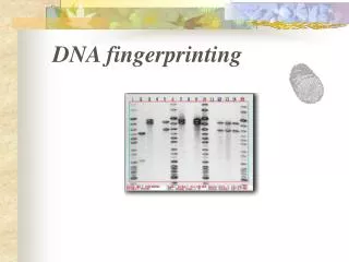
Results! • DNA fragments of the same size will form a band in the gel. DNA digested with an RE will show multiple bands on the gel. • A size standard (ladder) contains DNA fragments of known size…fragments in the sample DNA are compared to the ladder to estimate their size • The pattern of DNA fragments on a gel may be used to compare different samples
Electrophoresis Review • electrophoresis review
HYBRIDIZATION • This technique, also called blotting, is like finding a needle in a haystack. • Needle = DNA fragment of interest • Haystack = all the DNA fragments on a gel
Introduction • Hybrid DNA: artificially created double- stranded DNA molecule where each strand is from a different source. • Based on complementary base pairing rules of DNA • A always binds with T • C always binds with G
Types of Blotting • Southern Blotting was discovered by Edward Southern in 1975 • Other types of blotting have been discovered since then…their are a spoof on Southern’s naming his technique after himself. TypeSampleProbe Southern DNA DNA Northern RNA DNA Western protein protein antibody to first protein
Southern Blotting Components • RE digested DNA fragments separated on a gel • Basic (pH) solution to denature (break apart the two strands) the DNA • Membrane to which DNA fragments can bind • Single-stranded DNA probe • Short sequence of DNA that is complementary to the sequence you are searching for • Probe is labeled with a tag that makes it able to be seen • Radioactivity is often used as a tag • Visualization system for the probe • With radioactive probe, membrane is visualized with x-ray film
Blotting Steps • Denature the DNA by soaking the gel in a basic solution • Transfer single-stranded DNA to membrane by capillary action (movement of liquids against the forces of gravity due to surface tensions) • Incubate membrane with solution containing the tagged probe…probe anneals (binds) to its complementary sequence in the sample DNA • Wash off probe solution and visualize the blot
Review Southern Blotting • Southern blot demo • Shockwave required
Polymerase Chain Reaction • PCR is like a copy machine for a specific piece of DNA • Discovered by Kary Mullis is 1985 • Based on primer extension by DNA polymerase, discovered by Arthur Kornberg in 1960
DNA Replication in a Tube • DNA of interest, called the template • DNA primers anneal to the template • DNA polymerase synthesizes a complementary strand to the template by adding A, C G, or Ts where they are needed
PCR Components • Primers • Short strands of DNA, about 20 nt long • Nt sequence is complementary to each end of template DNA • Added in excess in PCR reaction so the primers will bind to the template DNA before the two longer template strands can anneal back to each other
PCR Components • Template DNA containing the region that you wish to amplify • dNTPs • Also called bases • n stands for A, C, G, and T…all 4 bases are included
PCR Components • Taq Polymerase • Heat stable DNA polymerase…otherwise you would need to add new DNA polymerase at the start of each cycle • Comes from the bacteria Thermus aquaticus, which live in hot springs and can withstand high temperatures
PCR Components • Thermalcycler • Heat block that can change temperatures rapidly • Controlled by a microcomputer • Incubation times & temperatures can be programmed in so thermalcycler runs automatically
PCR Steps • Mix components in tube and load into programmed thermalcycler • Denature at 94° C • H bonds broken so 2 strands of DNA come apart • Anneal between 50-60° C • Primer binds to complementary sequence on both strands • Synthesis at 72° C • Taq adds nts to elongate the 2 new strands in the 5’ 3’ direction
Copies Increase Exponentially • Each cycle doubles the # of copies of target sequence of DNA. • Theoretical yield will be 2n copies where n = number of PCR cycles • 30 cycles of PCR will yield > 1 billion copies!
PCR in Action pcr demo Shockwave required