Complex Cases of Malignant External Otitis and Necrotizing Otitis Externa in Elderly Patients
210 likes | 457 Vues
Detailed case studies of two elderly patients with severe external otitis, including symptoms, imaging findings, treatments, and challenges faced. Recommendations and therapeutic interventions discussed.

Complex Cases of Malignant External Otitis and Necrotizing Otitis Externa in Elderly Patients
E N D
Presentation Transcript
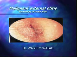
Malignant external otitis Necrotizing external otitis Dr. WASEEM WATAD
Case 1. ( SH. Y ) • 80 years old • 3VD , PTCA , DM-type2 , HTN , BPH • Ext. otitis with PO ABX and ear drops with improvement several months before admission • severe Rt. otalgia , facial pain Rt. , and Rt. parotid mass at admission 19/09/04 • Rt ear discharge • Weight loss
Case 1. • CT scan (20/09/04): Rt parotid mass , infiltration of parapharyngeal fat , EAC , infratemporal fossa , Rt. lat. pterygoid and masseter .no bony erosion and no lymphadenopathy • MRI (19/10/04) :process infiltrating the Rt. ear,temporal bone , TMJ, sphenoid sinus , infratemporal fossa and skull base • Biopsy of EAC polyp, parotid FNA (28/10/04) – mixed inflammation • Positive culture for p. aeruginosa
Case 1. • IV ABX treatment ( cephalosporine and quinolones ) with ear drops and toilette • Improvement in pain , ear discharge • There was no CN involvement
Case 2. ( Va. D ) • 68 years old • DM-type 2 , HTN • Hyperlipidemia , s/p CVA • Rt. Nasopharyngeal mass – biopsy no malignancy (11/04) • Bil. Ext. otitis 09/04 ( several weeks before admittion ) prolong ABX treatment ( semi-synthetic penicillin , quinolone) and ear drops
Case 2. • No improvement • Rt. Severe otalgia , ear discharge , persistent rt. ext. otitis , with granulation tissue • Elevated ESR , negative culture for p. aeruginosa • Start IV ceftazidime ( 5 weeks ) • Progression findings in serial CT/MRI
Case 2. • CT scan ( 14/11/04 ) - infiltration of the rt. parapharyngeal space , rt. Mastoid and middle ear, infiltrating of infratemporal fossa • MRI ( 24/21/04 ) – large mass in rt. parapharyngeal space with involvement of rt. TMJ and deep lobe of rt. Parotis • CT (01/05) infiltrating in rt. TMJ
Case 2. • De’bridment - (10/01/05) ,. (24/01/05), • Hx – inflammatory tissue • 2 weeks of AMIKACIN + MEROPENEM • Exacerbation of Rt. Otalgia , ear discharge and relapse of granulation tissue of EAC • Treatment failure ?? • Further therapy : • Broad spectrum of ABX – combination of cephalosporines and quinolone • Surgical treatment – mastoidectomy +/- tympanoplasty , ablation of granulating and necrotizing tissue, bone and cartilage sequestrations • HBO
Parietal Frontal Temporal Sphenoid Z Maxilla Lat. Pterygoid Plate Pterygomaxillary Fissure Infratemporal Fossa
MEO - criteria • Sade’ (1989) : • Severe EXT. otitis unresponsive to at least 10 days of conservative treatment • Increasing agonizing pain exacerbated at night • Granulation tissue in the base of EAC • Repeated isolation of pseudomonas • Levenson (1991) : • Refractory otitis ext. • Severe otalgia , worse at night • Purulent exudate , granulation tissue • Recovery of P. aeruginosa • DM , immune state compromise • Positive Tc-99 bone scan of temporal bone
MEO - staging • Corey (1985) : • I - Infection of bone and soft tissue without cranial nerves lesions or intracranial lesions • II- cranial nerve paralysis • a- VII paralysis only • b- Multiple cranial nerves paralysis • III – meningitis , epidural empyema , subdural empyema or brain abscess
NEO - diagnosis • Clinical findings • Laboratory tests • Culture • Ga-67, Tc-99 scans • HR-CT with contrast • Biopsy of granulation tissue
mortality • 46% (1968) • 10% recent articles • High mortality in facial n. paralysis
Management – cont. • HR-CT contrast evaluation • Ga-67 (every 4 weeks) follow up with treatment • Management underlying process ( DM / immunosuppressive) • Surgical de’bridment ,drinage – intracranial ext. , brain abscess • 6 weeks of ABX , repeat cultures , oral ABX after 2 weeks of cessation of symptoms
Management – cont. • Deep biopsy of granulation tissue – underlying carcinoma
Therapeutic problems • Main problem is : • Choice of the ABX • Duration of treatment
Therapeutic problems • Duration of treatment • Standard indication ( 6-8 weeks ) • Identifying objective parameter of definitive recovery • Healing of skin EAC • ESR • Ga-67
Therapeutic problems • Surgical treatment : • Complementary role • Mastoidectomy +/- tympanoplasty • Recommendation – biopsy , cleansing , ablation of necrotizing and granulation tissue and the bone , cartilage sequestrations
Therapeutic problems • Hyperbaric oxygen therapy • Daily , 2.4-3 atm, 90 minutea , 30 courses • Indications : advanced stages , recurrent cases, refractory to ABX • Hypoxia impaired oxygen dependent bacterial killing by phagocytosis