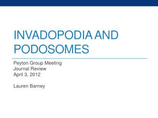
Invadopodia and Podosomes
500 likes | 1.22k Vues
Invadopodia and Podosomes. Peyton Group Meeting Journal Review April 3, 2012 Lauren Barney. Migration. Migration is a very important process in vivo Necessary for immune responses, wound healing, and embryonic development

Invadopodia and Podosomes
E N D
Presentation Transcript
Invadopodia and Podosomes Peyton Group Meeting Journal Review April 3, 2012 Lauren Barney
Migration • Migration is a very important process in vivo • Necessary for immune responses, wound healing, and embryonic development • Uncontrolled cell migration occurs in cardiovascular, immune, and developmental diseases as well as in tumor formation and metastasis • Barriers in the body can prevent cell migration unless the cells can get through them Bravo-Cordero, et al. (2012)
Invasion • Cells often must invade to cross membranes or barriers in the body • Boundaries: blood vessel walls, ECM, tissue boundaries • Invadosomesincluding invadopodiaand podosomes are cell-ECM contacts that are used when cells need to cross boundaries • Examples: • Cancer metastasis • Migration of immune cells to sites of infection Bravo-Cordero, et al. (2012)
What are invadopodia and podosomes? • Actin rich protrusions used for adhesion to and degradation of the ECM • They are significantly different than focal adhesions in structure and function • Used for invasion through the matrix or for crossing barriers in the body • Some cell types have an intrinsic ability to form invadopodia or podosomes, and sometimes they can be induced with growth factors or other stimulation Gray circles: viniculin-rich adhesion site Gimona, Grashoffand Kopp(2005)
Types of Cell Adhesion Structures Murphy and Courtneidge (2011)
Clinical Relevance Rous sarcoma virus –transformed rosette Linder, Wiesnerand Himmel (2011)
What are the differences between invadopodia and podosomes? Invadopodia Podosomes Shorter lived (on the order of minutes) Podosomes can form a ring structure called a rosette There is a ring around the actin core containing adhesion proteins such as vinculin or talin Often at the leading edge Cells often have many podosomes (10-100) Size: 1μm diameter, 0.4 μmdeep • Longer lived (on the order of an hour or more) • Are typically separate from one another, but can appear in groups • No ring of proteins around the actin core • Often around the nucleus or the leading edge • Generally, cells with invadopodia have 1-10 • Size: 8μm diameter, 5 μmdeep
Structure of Invadopodia and Podosomes Invadopodia Podosomes Linder, Wiesnerand Himmel (2011)
Imaging • C: Podosomes (yellow) in smooth muscle cells • D: Podosome rosettes (yellow) in epithelial cells • G: Invadopodia (red) near the center of a cell on a green fluorescent ECM substrate Gimona, Grashoff, and Kopp(2005)
How do invadopodia and podosomes degrade the ECM? • They secrete matrix metalloproteinases(MMPs) • A family of proteins which can degrade proteins • Many are secreted, but can be membrane proteins • Can secrete other proteases to degrade the ECM proteins • Initiation of invadopodia or podosomes creates a migratory phenotype, where focal adhesions are dissolved and the cell can begin to migrate Linder (2007)
What can induce the formation of invadopodia and podosomes? • Growth factors: result in phosphorylation of key proteins in the pathway • Platelet derived growth factor (PDGF) • Transforming growth factor-β (TGFβ) • Epidermal growth factor (EGF) • Integrins • αvβ3 integrin: found in podosomes of osteoclasts and invadopodia of several types of cancer • Interference with the αvβ3 integrin results in defective podosomes • The β1 integrin subunit is also found in both podosomes and invadopodia • Activation of the β1 integrin subunit induces invadopodia formation • Stiffness and other environmental cues
Stiffness Effects • Both invadopodia and podosomes have recently been shown to be mechanosensors, which can use stiffness cues to induce ECM degradation and migration • Stiffness has been shown to influence the lifetime and spacing of podosomes • Rigidity is important in matrix degradation with invadopodia • There is an optimal rigidity for invadpodia formation and ECM degradation (~30 kPa) • Invadopodia and podosomes can also be used for signal transduction within the cell via mechanotransduction
Assembly and Maturation of Invadopodia Murphy and Courtneidge (2011)
Invadopodia can be induced by growth factors or integrins http://www.latrobe.edu.au/biochemistry/lab/lock/index.htm
Cortactin • Protein in the cytoplasm of cells which can become activated by stimuli to promote rearrangement of the actin cytoskeleton • Involved in the formation of lamellipodia, invadopodia, as well as cell migration and endocytosis • Becomes activated in invadopodia; phosphorolated on three tyrosine residues • Has been shown to regulate the secretion of MMPs at invadopodia Cosen-Binker and Kapus (2006)
Immunofluorescent Imaging • Invadopodia are usually quantified using IF imaging • Actin is localized into punctate spots at invadopodia and podosomes • Also stain for other specific invadopodia or podosome markers • Cortactin: although, it also can randomly colocailize with actin, so confocal microscopy must be used to ensure that it is the bottom of the cell • TKS5: a key adaptor protein in invadopodia and podosomes which is not present in focal adhesions • Expression of TKS5 actually induces the formation of invadopodia, even in cells which don’t normally have them • Colocalization of actin with an invadopodia marker and local ECM degradation • See a void in a fluorescent ECM matrix that is colocalized with punctate spots of actin and the invadopodia marker (cortactin, etc)
Imaging and Quantification of Invadopodia • MTLn3 cells derived from the 13762NF rat mammary adenocarcinoma • C: cells have invadopodia until they are treated with an EGF receptor kinase inhibitor • D: serum starved cells don’t have invadopodia, but invadopodia form when cells are treated with EGF Yamaguchi, Pixleyand Condeelis (2006)
Fluorescent Matrix Bowden, et al. (2006)
Scanning Electron Microscopy • An invadopodium on a MDA-MB-231 cell seeded on pore filters Linder, Wiesnerand Himmel (2011)
Conclusions • This is a still very recent field; podosomes were first seen in 1985 and invadopodia were first described in 1994 • Currently, the classification of invadopodia and podosomes has not been clearly defined • It is also not clear whether invadopodia and podosomes originate from the same basic structures, even though they have similar pathways, which also haven’t been identified completely • However, an understanding of roles of invadopodia and podosomes in cell migration is crucial for making improvements in understanding and treating diseases
References • Albiges-Rizo, et al. “Actin machinery and mechanosensitivity in invadopodia, podosomes and focal adhesions.” Journal of Cell Science122(2009) 3037-3049 • Bowden, et al. “Co-localization of cortactin and phosphotyrosine identifies active invadopodia in human breast cancer cells.” Experimental Cell Research312 (2006) 1240-1253 • Bravo-Cordero, et al. “Directed cell invasion and migration during metastasis,” CurrOpin Cell Biol(2012) • Cosen-Binker and Kapus. “Cortactin: The Gray Eminence of the Cytoskeleton.” Physiology21 (2006) 352-361 • Gimona, Grashoff and Kopp. “Oktoberfest for adhesion structures.” MBO reports (2005) 6, 922–926. • Linder, “The matrix corroded: podosomes and invadopodia in extracellular matrix degradation”Trends in Cell Biology 17 (2007) • Linder, Wiesnerand Himmel, “Degrading Devices: Invadosomes in Proteolytic Cell Invasion.” Annu. Rev. Cell Dev. Biol. 27 (2011) 185–211. • Murphy and Courtneidge, “The ‘ins’ and ‘outs’ of podosomes and invadopodia: characteristics, formation and function.” Nature Reviews Molecular Cell Biology 12 (2011) 413-426 • Stylli, Kaye and Lock. “Invadopodia: At the cutting edge of tumourinvasion.” Journal of Clinical Neuroscience 15 (2008) 725–737 • Yamaguchi, Pixley and Condeelis. “Invadopodia and podosomes in tumor invasion.” European Journal of Cell Biology 85 (2006) 231-218.