Elective Project
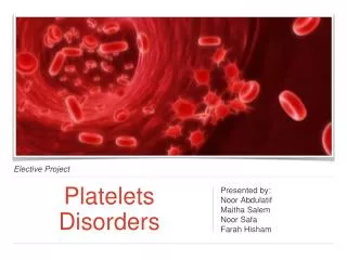
Elective Project. Platelets Disorders. Presented by: Noor Abdulatif Maitha Salem Noor Safa Farah Hisham. Objectives. Events of hemostasis Mechanism of vasoconstriction, platelets plug, blood coagulation. Structure & function of platelets Normal blood count and causes of variation

Elective Project
E N D
Presentation Transcript
Elective Project • Platelets Disorders • Presented by: • Noor Abdulatif • Maitha Salem • Noor Safa • Farah Hisham
Objectives • Events of hemostasis • Mechanism of vasoconstriction, platelets plug, blood coagulation. • Structure & function of platelets • Normal blood count and causes of variation • Blood coagulation tests • Platelets disorders • Disorder in number. • Disorder in function. • Deferential diagnosis of bleeding disorders.
Hemostasis Response to Injury
Platelets • Platelets (thrombocytes) are fragment of the cytoplasm of megakaryocyte. • They are small, colourless, andnon nucleated cells. • Shape: spherical or rod shaped • Size: 1 to 4 μmindiameter. • Life span: 8 – 10 days • Normal count: 150,000 to 450,000 / μl
Phospholipids, relaese of clotting factors,and Ca2+ Functions of Platelets • Glycoprotein on the surface coat that adhere it to injured endothelial cells. • Prevent • Bleeding • Blood • Clotting
Functions of Platelets • Secretes growth factor PDGF that promotes growth & multiplication of vascular endothelial cells • Actin, myosin & thrombosthenin • that are contractile proteins • Clot • Retraction • Repair damaged vascular wall
Hemostasis - Plug Formation Formation of platelet plug:
Hemostasis - Plug Formation Platelet Adhesion :
Hemostasis - Plug Formation Platelet Activation :
Hemostasis - Plug Formation Platelets aggregation :
Hemostasis - Blood Coagulation Conversion of Fibrinogen to Fibrin :
Hemostasis Prevention of Blood Clotting in the Normal Vascular System—Intravascular Anticoagulants The smoothness of the endothelial cell surface, which prevents contact activation of the intrinsic clotting system; A layer of glycocalyx on the endothelium (glycocalyx is a mucopolysaccharide adsorbed to the surfaces of the endothelial cells), which repels clotting factors and platelets, thereby preventing activation of clotting; and An alphaglobulin called antithrombin III or antithrombin-heparin cofactor. Heparin
Hemostasis A protein bound with the endothelial membrane, thrombomodulin, which binds thrombin ▼ Prevent clotting in normal vascular system BUT HOW?
Hemostasis – Lysis of Blood Clots • The plasma proteins contain an euglobulin called plasminogen (or profibrinolysin) that, when activated, becomes a substance called plasmin (or fibrinolysin). • Plasmin digests fibrin fibers and some other protein coagulants such as fibrinogen, Factor V, Factor VIII, prothrombin, and Factor XII. • It can cause lysis of a clot by destroying many of the clotting factors, thereby sometimes even causing hypocoagulability of the blood.
Hemostasis – Lysis of Blood Clots • The injured tissues and vascular endothelium very slowly release a powerful activator called tissue plasminogen activator (t-PA) that a few days later, after the clot has stopped the bleeding. • Converts plasminogen to plasmin, which in turn removes the remaining unnecessary blood clot.
Assesment of hemostatic functions • Bleeding time. • Clotting time. • Prothrombin time and international normalized ratio. • Activated Partial Thromboplastin Time test
Bleeding time • Bleeding usually stops within 1 to 6 minutes. • Lack of any one of the several of the clotting factors can prolong the bleeding time, but it is especially prolonged by lack of platelets.
Clotting time • Normal clotting time: 6 to 10 minutes. • No longer used in many clinics.
Prothrombin Time • How it is performed: • Adding the patient's plasma to some source of Tissue Factor and Calcium chloride. • PTT measures: • The integrity of the extrinsic system as well as factors common to both systems. • Normal PT Values: 10-12 seconds.
International Normalized Ratio The INR is used to make sure the results from a PT test is the same at one lab as it is at another lab. Normal INR Values: 1 to 2
Activated Partial ThromboplastinTime • The test is performed to: • Determine if heparin therapy is effective. • Detect the presence of a clotting disorder. • How it is performed: • Calcium and activating substances (kaolin and cephalin) are added. • PTT measures: • The integrity of the intrinsic system (Factors XII, XI, VIII, IX) and common clotting pathways. • Normal PTT Values: 30 to 45 seconds
Causes of abnormal hemostasis Causes of abnormal hemostasis: • Platelets disorders. • Coagulation disorders. • Blood vessel wall disorder. • Fibrinolysisdisorders.
Thrombocytosis • Thrombocytopenia • Acquired • Inherited • Disorders in Number • Disorders in Function • Platelets Disorders
Causes of variation in platelets count • A normal platelet count in a healthy individual is between 150,000 and 450,000 / μl of blood. • Thrombocytopenia is the term used for cases having low platelets count. • Thrombocytosis is the term used for cases having high platelets count. • An abnormality or disease of the platelets is called a thrombocytopathy.
Disorders in Number - Thrombocytosis Thrombocytosis: • It is a disorder in which the body produces too many platelets (> 500,000\μl) • When it’s caused by a blood and bone marrow disease, it is called essential/primary thrombocythemia. • When it's caused by an underlying condition, it is called reactive/secondary thrombocytosis.
Disorders in Number - Thrombocytosis Essential Thrombocythemia • What causes this to happen usually is unknown. • Faulty stem cells in the bone marrow make too many platelets. • The platelets are abnormal in primary thrombocythemia. • A rare form of thrombocythemia is inherited.
Disorders in Number - Thrombocytosis Reactive Thrombocytosis • Some of the conditions or factors that can cause a high platelet count are: • Cancer • Iron-deficiency anemia. • Acute hemolytic anemia. • Splenectomy. • Inflammatory or infectious diseases.
Disorders in Number - Thrombocytosis Symptoms & Signs • People who have primary thrombocythemia are more likely than those who have secondary thrombocytosis to have serious signs and symptoms. • They include: • Weakness • Thrombosis • Headache • Dizziness • Chest pain • Numbness in the hands and feet.
Disorders in Number - Thrombocytosis Treatment • Essential thrombocythemia: • Medications: - Hydroxyurea. • Plateleltpheresis • Reactive thrombocytosis istreated by treating the underlying cause.
Disorders in Number - Thrombocytopenia Thrombocytopenia • Presence of low numbers of platelets (<150,000/μl) in the circulating blood. • Classification: • Mild (100,000-150,000/μl) • Moderate (50,000-100,000/μl) • Severe (<50,000/μl) • Serious ( <5000/μl) • Bleeding is usually from many small venules or capillaries. • The skin displays many small, purplish blotches, giving the disease the name thrombocytopenic purpura.
Disorders in Number - Thrombocytopenia Causes Production failure: • Bone marrow diseases. Consumptive disorder (the platelets are destroyed in the peripheral circulation & spleen): • Autoimmune diseases (ITP). • Infections. • Splenomegaly. • Medicines: - Heparin - Some antibiotics
Disorders in Number - Thrombocytopenia Symptoms & Signs External bleeding: - Purpura and petechiae - Nosebleed - Prolonged bleeding from minor cuts Internal bleeding: - Blood in the urine or stool
Purpura • Purpura: bleeding under the skin or into mucosal membranes. • Purpura can be subdivided based on size into petechiae and ecchymoses. • Pinpoint areas (less than 2 mm) of hemorrhage, which are reddish-purple lesions are called petechiae. • Larger confluent lesions are referred to as ecchymoses. • Ecchymoses are commonly called bruises.
Disorders in Number - Thrombocytopenia Treatment • Medicines: • Steroids. • IV immunoglobulins. • Anti-Rh antibody. • Blood or platelets transfusion. • Splenectomy. • Immunosuppressnt agent.
Thrombocytosis • Thrombocytopenia • Acquired • Inherited • Disorders in Number • Disorders in Function • Platelets Disorders
Disorders in Function –Bernard-Soulier Syndrome • An autosomal recessive disorder • Deficiency on the surface of the platelet in Glycoprotein Ib/IX. • Platelets fail to stick and clump together at the site of the injury. • Bleeding time is prolonged, and platelets count is low.