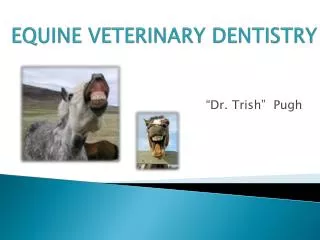
Preventative Veterinary Dentistry
Preventative Veterinary Dentistry. How to, charts and home care. Introduction. You do not need to write this. By the end of this power point, you should be able to: Understand how to complete an oral examination Know the basic dental procedures for operative cases

Preventative Veterinary Dentistry
E N D
Presentation Transcript
Preventative Veterinary Dentistry How to, charts and home care
Introduction You do not need to write this • By the end of this power point, you should be able to: • Understand how to complete an oral examination • Know the basic dental procedures for operative cases • Carry out a charting procedure • Understand the need to initiate and sustain dental homecare
Oral Examination • Essential as part of the overall physical exam • Domestic pets are substantially less demonstrative of oral pain then humans • Because of this, owners may be unaware of major dental problems—makes the oral examination more important!
Taking the History • This is the first step of any exam. • You need to determine the following: • Age • Breed • Sex • Environmental Concerns* • Presenting Signs* • Oral History* • *on next slides
Environmental Concerns • Vaccinal status • Diet • Possible behavior changes • Home environment: yard or house? • Pet or working animal?
Presenting Signs (symptoms) • Possible weight loss • Anorexia • Halitosis (Bad Breath!) • Dysphagia (Difficulty Swallowing) • Abnormal Salivation • Retching or vomiting • Chattering Jaws • Inability or difficulty to close/open mouth • Facial swelling or disfigurement
Oral History • Previous dental procedures? • Charts? • Present and previous level of homecare?
Oral Exam Procedures • 1. Visually inspect head and neck from afar. Palpate outer surfaces of head for pain, heat, sensitivity or swelling. Palpate lymph nodes • 2. Inspect lips and palpate. Retract lips and examine vestibule (inner surfaces) • 3. Open the mouth (checking for pain)
4. Examine the mucous membranes. Check for color, inflammation, ulceration, hyperplasia, bleeding, unusual swelling, tumors and foreign bodies. • 5. Examine teeth for calculus, gingivitis, malocclusion of bite, absence or broken teeth, and enamel abnormalities.
Diagnostic Techniques • Functional studies: • Offer the patient food and/or water. Owner’s might not be able to give you enough information. • Imaging: • This is the #1 tool for diagnosis! • CT Scans and MRI’s are becoming more available for inaccessible areas such as TMJ’s, sinuses and nasal cavity.
Biopsy: • Essential for lesions in the mouth (a lot present the same) • Called a biopsy punch (takes sample of tissue) • Culture: • Can be of help, but is over-rated. • Will detect what is present (bacteria) but not why, so more tests will be needed.
Dental Charting!! • This is the FIRST STEP in basic dental procedures!! • WHY chart? • Records • Success of treatments is impossible to gauge over time without tracking • It’s a good clinical habit to develop
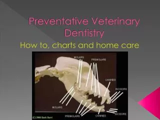
DOG CHART: • Note the graphics (on your chart) • Top line indicates the buccal (lateral) view. Lower line indicates the occlusal view—seen as if you were inside the mouth looking out. • 3 boxes for the upper and lower jaws can be used for 3 separate dental procedures. Allows for monitoring over time. • Some findings may be best drawn onto the teeth (periodontal pockets, caries*, fractures, etc.) • *caries: tooth decay
CAT CHART: • Basically the same as the dog but less teeth (30 rather than 42)
Sequence of Charting • 1. Count the teeth and note missing or extra teeth • 2. Determine level of calculus as per the legend • 3. Determine level of gingival inflammation as per the legend • 4. Note any major visible abnormalities—fractured teeth, enamel defects, tooth decay, etc.
5. Note the location of any teeth extracted by crossing out the corresponding boxes and tooth graphics • 6. Note any other impt features such as: • Gingival recession and root exposure • Caries—draw location • Enamel defects—draw location • Mobile teeth—use legend • Other significant problems • 7. Note treatments performed—fillings, extractions, root canals, etc.
Scaling and Polishing(You need to find these for homework and explain them!) • 1. Removing calculus • 2. Explore and Probe • 3. Subgingival Scaling • 4. Polishing • 5. Irrigation • 6. Fluoride or Barrier Treatment
HOMECARE!! • The MOST IMPT aspect of a basic dental procedure!! • Must review with the owner: • How to brush teeth • It is a DAILY procedure • Products to use (brushes, toothpaste, treats and toys to help)
Key Points • 1. A thorough oral examination with history is essential to enable you to relate the findings to the body as a whole. • 2. Charting the mouth to record the findings before and after surgery is essential to keep track of the case. • 3. The homecare program is the most impt part of prevention. It should be constructed, initiated and monitored with relevance to the needs of the patient and ability of the owner.
References • 2002 eMedia Unit RVC