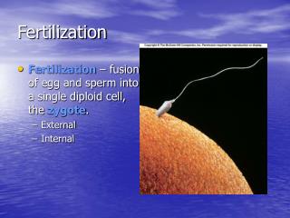
The Journey of Fertilization: Barriers, Capacitation, and Gamete Fusion
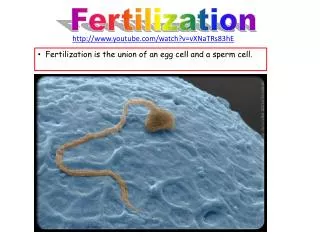
Understand the intricate process of fertilization, from sperm transport to fusion with the ovum. Explore barriers sperm face, capacitation stages, acrosome reaction, and the crucial block to polyspermy. Discover the fascinating journey of creating new life.

The Journey of Fertilization: Barriers, Capacitation, and Gamete Fusion
E N D
Presentation Transcript
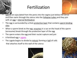
Sperm transport Semen is ejaculated into vagina, cervical os and may reach cervical canal Semen coagulate by coagulating enzymes from prostate gland which interacts with fibrinogenous substrate from seminal vesicles Coagulum protects spermatozoa from vaginal acidity and prevent loss of spermatozoa Coagulum liquefies after 15 to 20 minutes Liquefaction time is one criteria for semen quality Sperm may live up to 48 – 72 hours in female reproductive tract
Barriers (1) Spermatozoa have to confront three barriers before reaching ampulla, the site for fertilization First barrier – cervical mucus Mucus will filter and choose spermatoza – dead and immotile spermatozoa are discarded, normal spermatozoa are stored in the cervical crypts (first reservoir) Filtered spermatozoa discarded in the vaginal secretion post-coital Consistency of cervical mucus assist in sperm motility upwards Mucus thick, sperm could not penetrate Mucus thin and more stringy, sperm is assisted in motility by swimming in channels form by the mucus
Barriers (2) Endometrial glands – second barrier Will choose and filter spermatozoa Chosen spermatozoa are stored here (second reservoir) Here, capacitation occurs due to prostaglandins from endometrium
Barriers (3) Third barrier – utero-tubal junction Chosen spermatozoa are finally stored in the isthmus (third reservoir) to wait for the ovum to travel down Ovulated ovum will be caught by infundibulum when fimbriae comes close to ovary Ovum will travel down the infundibulum to the ampulla by oviductal contraction and presence of cilia
Capacitation (1) A time dependent phenomenon which is species-specific Takes more than 24 hours in human Reversible process (if capacitated spermatozoa are placed in epididymal fluid or seminal plasma, will be decapacitated – contains decapacitating factor) only in vitro Must occur to enable acrosome reaction to occur Substances like cholesterol, glycosaminoglycans and glycoproteins are stripped from plasma membrane of sperm head
Capacitation (2) Two elements in this process: Hyperactivated motility – sperm starts to show whiplashing movement to enable sperm to move forward faster Change to membrane surface – membrane stability decreases. More permeable to calcium ions. Tyrosine kinase activity increases. Adenyl cyclase activity in spermatozoa heightens and causes protein phosphorylation which are cAMP dependent
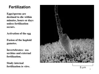
Acrosome reaction Occurs right after capacitation Totally dependent on calcium uptake into cells and increase in intracellular pH (pH 7.1 to pH 7.5) The acrosome swells and the outer acrosomal membrane fuses with the overlying plasma membrane Vesiculation occurs and pores are formed Acrosomal contents (hyaluronidase, acrosin etc) are released
Acrosome reaction Two types: True acrosome reaction – reaction occurs at zona pellucida False acrosome reaction – degeneration of sperm due to death (enzymes from acrosome will self-desctruct sperm)
Initiators of the acrosome reaction (1) High calcium influx ZP3 (zona pellucida glycoprotein 3) Progesterone etc ZP3 in ovum will bind to sperm binding protein (receptor) on sperm plasma membrane This binding site may contain galactosyl transferase activity When binding occurs, G protein involvement will stimulate calcium influx and the rise in pH initiates the acrosome reaction
Initiators of the acrosome reaction (2) Progesterone will also stimulate calcium influx which then stimulates adenyl cyclase and cAMP Progesterone can stimulate acrosomal leakage to release hyaluronidase Hyaluronidase will digest hyaluronic acid which binds cumulus cells When these cells breaks apart, spermatozoa can bind to zona pellucida Progesterone has been reported to initiate capacitation also
Sperm binding properties to zona Outer acrosomal membrane has receptor to ZP3 Inner acrosomal membranes has receptor to ZP2 Equatorial segment and post-acrosomal region is the part of the spermatozoon that enters the ovum Tail and midpiece left outside ovum
ZP3 binding with receptor on outer acrosomal membrane of sperm
ZP2 binding to receptor on inner acrosomal membrane of sperm
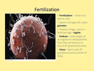
Gamete fusion Sperm penetration of the zona takes between 5-20 minutes Sperm lies tangent at the ovum surface between the zona pellucida and oolemma at the perivitelline space Microvilli at the oocyte surface will engulf the sperm head The equatorial segment and the post-acrosomal region of the sperm fuses with the ovum After fusion, zygote forms and male and female pronuclei Syngamy occurs when both male and female chromosomes combines and then form 2-cell conceptus
Block to Polyspermy Important to prevent more than one sperm fertilising the ovum. This phenomenom ensures: Prevention of triploidy (2 sperm fertilising an ovum) Prevention of polyploidy (many sperm fertilising an ovum) Usually only one sperm (n) can fertilize an ovum (n) to achieve a diploid (2n) zygote Fusion of ovum to sperm initiates the release of free intracellular calcium from calcium stores and this causes calcium spikes.
Increase in calcium is due to change in the pattern of protein phosphorylation in the ovum and the involvement of enzymes e.g., tyrosine kinase and phospholipase B High calcium influences the cortical reaction; cortical granules in the zygote cortex will fuse with the oolema. This causes release of the cortical substances into the cortical space The substances (enzymes) will act on the zona pellucida by ‘hardening’ it This hardening (zona pellucida reaction (tindakbalas zona) is the ‘masking’ of the ZP3 and ZP2 so that other sperm could not digest the zona with acrosin and also reduce the sperm binding properties to the zona
Mammalian sperm factor – PLC-zeta Schematic representation of the basic hypothesis for how PLC initiates Ca2+ release in mammalian eggs.
Stimulus of oocyte activation After fusion of the sperm and egg plasma membranes, the sperm-derived PLC protein diffuses into the egg cytoplasm. This hydrolyses PIP2, from an unknown source, to generate InsP3. It is possible that the subsequent rise in Ca2+ leads to the regulation of PLC activity.
When sperm activate eggs at fertilization the signal for activation involves increases in the intracellular free Ca2+ concentration. In mammals the Ca2+ changes at fertilization consist of intracellular Ca2+ oscillations that are driven by the generation of inositol 1,4,5-trisphosphate (InsP3). It is not established how sperm trigger the increases in InsP3 and Ca2+ at fertilization.
One theory suggests that sperm initiate signals to activate the egg by introducing a specific factor into the egg cytoplasm after membrane fusion. This theory has been mainly based upon the observation that injecting a cytosolic sperm protein factor into eggs can trigger the same pattern of Ca2+ oscillations induced by the sperm. The soluble sperm factor protein is a novel form of phospholipase C (PLC), and it is referred to as PLC (zeta).
Embryonic development (1) Germinal period (movement of zygote and implantation in uterus) lasts two weeks Cleavage occurs - 1 cell to 2 daughter cells after 36 hours post fertilization Daughter cells called blastomeres Zygote still covered by ZP ZP inhibits blastomeres from falling apart If this happens, two possibilities occur Monozygotic twins Chimaeras Chimaeras is the fusion of two different zygotes from two fertilized ovum – two sets of two different genotype
Fertilized egg 2 cell stage 4cell stage 8 cell stage
Embryonic development (2) Blastomeres becomes morula on day 3 Progesterone from functioning CL will stimulate the release of glycogen from endometrium for energy to developing embryo (histiotropic nutrition) High levels of progesterone also inhibit oviductal constriction to enable morula to move towards the uterus by peristaltic contraction and cilia movement Becomes blastocyst on day 4 - 5
Embryonic development (3) Blastocysyt has fluid-filled cavity (blastocoele) Has inner cell mass (ICM) surrounded by trophodectum (trophoblast) ICM will form extra embryonic membranes (amnion, yolk sac etc) and fetus Trophoblast forms chorion Blastocyst floats in uterine cavity for 1 – 2 days Prior to implantation, will shed ZP by enzymatic digestion
Maternal Recognition of Pregnancy Embryo sends signal along ovarian-pituitary pathway Stop FSH release but maintain LH Important because luteal regression will occur if the mother does not recognise the pregnancy Drop in progesterone will cause abortion Signal to mother is to change cyclic pattern of oscillating progesterone and estrogen to non-oscillating pattern with only progesterone as dominating hormone to maintain pregnancy Prostaglandin F2 secretion are also inhibited throughout pregnancy
Implantation (1) A nutritional and physical contact between fetus and mother Blastocyst surface becomes sticky Trophoblastic cells (cytotrophoblast) releases enzymes to digest proteins on endometrium Syncytiotrophoblast enters endometrium to suck up metabolic fuel and nutrients Deep invasion into endometrium occurs Change to endometrium occurs (stromal reaction/primary decidualization reaction) Endometrium releases prostaglandins to stimulate vascularization causing edema and increasing nutrient stores
Implantation (2) The invaded part of the endometrium is called decidua 2 –3 days post invasion, decidua enlarges to become secondary decidua Blastocyst enters this decidua After entry, a layer of endometrial cells will cover and bury the blastocyst Syncytiotrophoblast on the other hand keep on digesting endometrial cells to get nutrients until the placenta is formed