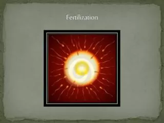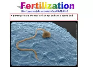

Fertilization
E N D
Presentation Transcript
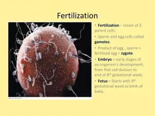
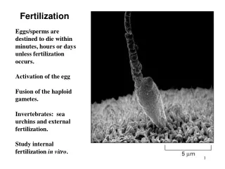
All the material necessary for the beginning of growth and development must be stored in the mature egg (the ovum). Whereas the sperm has eliminated most of its cytoplasm, the developing egg not only conserves its material, but is actively involved in accumulating more. The meiotic divisions that form the oocyte conserve its cytoplasm and the oocyte either synthesizes or absorbs proteins, such as yolk, that act as food reservoirs for the developing embryo. Thus, birds’ eggs are enormous single cells, swollen with their accumulated yolk. Even So, while sperm and egg have equal haploid nuclear components, the egg also has a remarkable cytoplasmic storehouse that it has accumulated during its maturation. This cytoplasm includes the following: • Proteins. The early embryonic cells need a supply of energy and amino acids.. Many of the yolk proteins are made in other organs (liver, fat body) and travel through the maternal blood to the egg. • Ribosomes and tRNA. The early embryo needs to make many of its own proteins, and in some species, there is a burst of protein synthesis soon after fertilization. Protein synthesis is accomplished by ribosomes and tRNA, which exist in the egg. • Messenger RNA. In most organisms, the instructions for proteins made during early development are already packaged in the oocyte. • Morphogenetic factors. They appear to be localized in different regions of the egg and become segregated into different cells during cleavage. • Protective chemicals. Many eggs contain ultraviolet filters and DNA repair enzymes that protect them from sunlight.
Eggs are protected by elaborate envelopes Vitelline envelope: a glycoprotein layer covers the plasma membrane of all eggs. This acts to protect the egg. Eggs that are deposited in water have a jelly-like coating that surrounds the egg (frogs eggs) Eggs that are deposited on land have particularly elaborate envelopes. The eggs of birds have a vitelline envelope, a fibrous layer, an outer layer of albumin (egg white), and a shell composed of calcium carbonate. The outer envelopes are synthesized in the oviduct after the egg has been fertilized.
Egg • Nucleus, cell membrane, vitelline envelope (zona pellucida), cortex and cortical granules, mitocondria, jelly cortical granules analogous to acrosome – contains enzymes, mucopolysaccharides, adhesive glycopoteins, etc
Different types of eggs • Term Yolk Cleavage Result Model Organisms Notes • Alecithal Little Holoblastic Blastocyst Mammal • Centrolecithal Center Superficial Blastoderm Insect • Isolecithal Even Holobastic Blastula Urchin, Sand Dollar • Mesolecithal Uneven Holoblastic Blastula Amphibian Sometime telolecithal. • Telolecithal Uneven Meroblastic Blastodisc Bird,fish,reptile Sometimesextreme telolecithal.
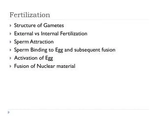
Egg • Stages of egg maturation at the time of sperm entry in different animal species. The germinal vesicle is the name given to the large diploid nucleus of the primary oocyte. The polar bodies are seen as smaller cells.
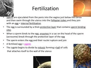
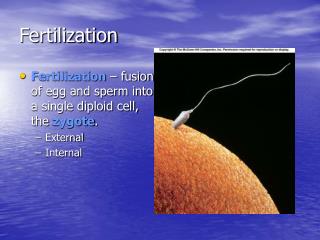
Types of fertilization Internal fertilization and external fertilization • Internal means When the fusion of the male and female gamete occurs within the female genital tract it is called internal fertilization. This type of fertilization occurs in all terrestrial animals both oviparous and viviparous. Reptiles, Birds and mammals show internal fertilization • The fusion of the gametes occurs outside the body of the animal and is common in aquatic animals. In bony fishes, frogs, echinoderms the sperms and ova are released into the water and their union occurs by Chromosome number of few chance. Since fertilization occurs outside the body of the animal externally, this method is called external fertilization.
Animal – vegetal polarity: In eggs that have a lot of yolk, the yolk is concentrated in the vegetal pole. The animal pole contains the nucleus and relatively little yolk. The yolk in the vegetal pole interferes with cytokinesis during the process of cleavage leading to incomplete cleavage. he eggs and zygotes of most animals, except mammals, have a definite polarity. The polarity is defined by the uneven distribution of mRNA, proteins, yolk (stored nutrients) etc. in the cytoplasm; this creates the animal and vegetal hemispheres. The yolk is concentrated in the vegetal pole of the egg and makes the pole yellow. The vegetal pole becomes the posterior of the embryos, while the animal pole becomes the anterior. At fertilization, the cortex of the animal hemisphere slides over the underlying cytoplasm towards the point of sperm entry. It exposes a region called the gray crescent opposite the point of sperm entry; the gray crescent marks the dorsal side of the future embryo. The first cleavage bisects the gray crescent and creates the left and right axes of the embryo.
Specification of the chick anterior-posterior axis by gravity. Rotation in the shell gland (A) results in the lighter components of the yolk pushing up one side of the blastoderm (B). That more elevated region becomes the posterior of the embryo
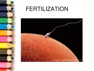
Recognition of Egg and Sperm • The chemoattraction of the sperm to the egg by soluble molecules secreted by the egg • The exocytosis of the acrosomal vesicle to release its enzymes • The binding of the sperm to the extracellular envelope (vitelline layer or zona pellucida) of the egg • The passing of the sperm through this extracellular envelope • Fusion of egg and sperm cell plasma membranes
Chemo taxis • In many species, sperm are attracted toward eggs of their species by chemotaxis, that is, by following a gradient of a chemical secreted by the egg. The mechanisms of chemotaxis differamong species. One chemotactic molecule, a 14-amino acid peptide called resact, has been isolated from the egg jelly of the sea urchin Arbacia punctulata. Resact diffuses readily in seawater and has a profound effect at very low concentrations when added to a suspension of Arbacia sperm
Sperm – Egg InteractionAcrosomal reaction • components of egg jelly bind to receptors on sperm cell membrane • calcium channels opened, calcium enters sperm head and induces fusion of acrosomal vesicle with membrane leading to exocytosis of enzymes • acrosomal process forms from polymerization of actin also facilitated by calcium
Species-specific recognition in sea urchins • Once the sea urchin sperm has penetrated the egg jelly, the acrosomal process of the sperm contacts the surface of the egg . A major species-specific recognition step occurs at this point. The acrosomal protein mediating this recognition is called bindin. In 1977, Vacquier and co-workers isolated this nonsoluble 30,500-Da protein from the acrosome of Strongylocentrotus purpuratus and found it to be capable of binding to dejellied eggs of the same species Further, its interaction with eggs is relatively species-specific bindin isolated from the acrosomes of S. purpuratus binds to its own dejellied eggs, but not to those of Arbacia punctulata.
Immunochemical technique used to localize bindin. Rabbit antibody was made to the bindin protein, and this antibody was incubated with sperm that had undergone the acrosomal reaction. If bindin was present, the rabbit antibody would remain bound to the sperm. After any unbound antibody was washed off, the sperm were treated with swine antibody that had been covalently linked to peroxidase enzymes. The swine antibody bound to the rabbit antibody, placing peroxidase molecules wherever bindin was present. Peroxidase catalyzes the formation of a dark precipitate from diaminobenzidine (DAB) and hydrogen peroxide. Thus, this precipitate formed only where bindin was present.
Gamete binding and recognition in mammals ZP3 glycoprotein of egg binds sperm and initiates acrosomal reaction by binding to receptors that activates G protein and opens calcium channels leading to exocytosis enzymes digest opening in zona, as sperm crosses zona loses ZP3 binding sites but ZP2 binding sites come into play
ZP3-binding proteins on the mouse sperm are located in the plasma membrane, overlying the acrosome. In this confocal image, a ZP3-binding protein is stained red by antibody immunofluorescence.
Induction of the mammalian acrosomal reaction by ZP3 • Unlike the sea urchin acrosomal reaction, the acrosomal reaction in mammals occurs only after the sperm has bound to the zona pellucida. The mouse sperm acrosomal reaction is induced by the crosslinking of ZP3 with the receptors for it on the sperm membrane.This crosslinking opens calcium channels to increase the concentration of calcium in the sperm. The mechanism by which ZP3 induces the opening of the calcium channels and the subsequent exocytosis of the acrosome remains controversial, but it may involve the receptor’s activating a cation channel (for sodium, potassium, or calcium), which would change the resting potential of the sperm plasma membrane. The calcium channels in the membrane would be sensitive to this change in membrane potential, allowing calcium to enter the sperm.
Secondary binding of sperm to the zona pellucida • During the acrosomal reaction, the anterior portion of the sperm plasma membrane is shed from the sperm. This region is where the ZP3-binding proteins are located, and yet the sperm must still remain bound to the zona in order to lyse a path through it. Acrosome-intact sperm will not bind to ZP2, acrosome-reacted sperm will. Moreover, antibodies against the ZP2 glycoprotein will not prevent the binding of acrosome-intact sperm to the zona, but will inhibit the attachment of acrosome-reacted sperm. The structure of the zona consists of repeating units of ZP3 and ZP2, occasionally crosslinked by ZP1. It appears that the acrosome-reacted sperm transfer their binding from ZP3 to the adjacent ZP2 molecules. After a mouse sperm has entered the egg, the egg cortical granules release their contents. One of the proteins released by these granules is a protease that specifically alters ZP2. This inhibits other acrosome-reacted sperm from moving closer toward the egg.
Sperm – Egg Interaction • fusion of sperm and egg membranes • polymerization of egg actin leads to formation of fertilization cone in sea urchin • in some species parts of egg membrane specialized to fuse with sperm, in sea urchins all regions are capable • fusion mediated by certain proteins – bindin in sea urchins has secondary role
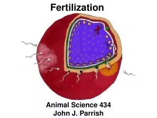
Gamete Fusion and the Prevention of Polyspermy • Sperm-egg binding appears to cause the extension of several microvilli to form the fertilization cone. The sperm and egg plasma membranes then join together, and material from the sperm membrane can later be found on the egg membrane. The sperm nucleus and tail pass through the resulting cytoplasmic bridge, which is widened by the actin polymerization. • A similar process occurs during the fusion of mammalian gametesIn mammals, the fertilin proteins in the sperm plasma membrane are essential for sperm membrane-egg membrane fusion Mouse fertilin is localized to the posterior plasma membrane of the sperm head). It adheres the sperm to the egg by binding to the α6β1 integrin protein on the egg plasma membrane. Moreover, like sea urchin bindin fertilin has a hydrophobic region that could potentially mediate the union of the two membranes. Thus, fertilin appears to bind the sperm plasma membrane to the egg plasma membrane and then to fuse the two of them together.
The prevention of polyspermy • The entrance of multiple sperm—polyspermy—leads to disastrous consequences in most organisms. In the sea urchin, fertilization by two sperm results in a triploid nucleus, in which each chromosome is represented three times rather than twice. Worse, since each sperm’s centriole divides to form the two poles of a mitotic apparatus, instead of a bipolar mitotic spindle separating the chromosomes into two cells, the triploid chromosomes may be divided into as many as four cells. Because there is no mechanism to ensure that each of the four cells receives the proper number and type of chromosomes, the chromosomes would be apportioned unequally. Some cells receive extra copies of certain chromosomes and other cells lack them.
The fast block to polyspermy • The fast block to poly-spermy is achieved by changing the electric potential of the egg plasma membrane. This membrane provides a selective barrier between the egg cytoplasm and the outside environment, and the ionic concentration of the egg differs greatly from that of its surroundings. This concentration difference is especially significant for sodium and potassium ions. Seawater has a particularly high sodium ion concentration, whereas the egg cytoplasm contains relatively little sodium. The reverse is the case with potassium ions. This condition is maintained by the plasma membrane, which steadfastly inhibits the entry of sodium ions into the oocyte and prevents potassium ions from leaking out into the environment. This resting membrane potential is generally about 70 mV, usually expressed as –70 mV because the inside of the cell is negatively charged with respect to the exterior.
The slow block to polyspermy • cortical granule reaction, a slower, mechanical block to polyspermy that becomes active about a minute after the first successful sperm-egg attachment. Directly beneath the sea urchin egg plasma membrane are about 15,000 cortical granules, each about 1 μm in diameter. Upon sperm entry, these cortical granules fuse with the egg plasma membrane and release their contents into the space between the plasma membrane and the fibrous mat of vitelline envelope proteins. Several proteins are released. The first are proteases. These enzymes dissolve the protein posts that connect the vitelline envelope proteins to the cell membrane, and they clip off the bindin receptor and any sperm attached to it .Mucopolysaccharides released by the cortical granules produce an osmotic gradient that causes water to rush into the space between the plasma membrane and the vitelline envelope, causing the envelope to expand and become the fertilization envelope. A third protein released by the cortical granules, a peroxidase enzyme, hardens the fertilization envelope by crosslinking tyrosine residues on adjacent proteins .The fertilization envelope starts to form at the site of sperm entry and continues its expansion around the egg. As it forms, bound sperm are released from the envelope. This process starts about 20 seconds after sperm attachment and is complete by the end of the first minute of fertilization. Finally, a fourth cortical granule protein, hyalin, forms a coating around the egg. The egg extends elongated microvilli whose tips attach to this hyaline layer. This layer provides support for the blastomeres during cleavage. In mammals, the cortical granule reaction does not create a fertilization envelope, but its ultimate effect is the same. Released enzymes modify the zona pellucida sperm receptors such that they can no longer bind sperm (Bl. During this process, called the zona reaction, both ZP3 and ZP2 are modified. in mice N-acetylglucosaminidase enzymes cleave it from ZP3
Calcium as the initiator of the cortical granule reaction • Upon fertilization, the intracellular calcium ion concentration of the egg increases greatly. In this high-calcium environment, the cortical granule membranes fuse with the egg plasma membrane, releasing their contents. Once the fusion of the cortical granules begins near the point of sperm entry, a wave of cortical granule exocytosis propagates around the cortex to the opposite side of the egg. • The calcium ions responsible for the cortical granule reaction are stored in the endoplasmic reticulum of the egg .In sea urchins and frogs, this reticulum is pronounced in the cortex and surrounds the cortical granules .In Xenopus, the cortical endoplasmic reticulum becomes ten times more abundant during the maturation of the egg and disappears locally within a minute after the wave of cortical granule exocytosis occurs in any region of the cortex. Once initiated, the release of calcium is self-propagating. Free calcium is able to release sequestered calcium from its storage sites, thus causing a wave of calcium ion release and cortical granule exocytosis.
Egg Activation • increase in calcium levels trigger many reactions • most protein synthesis comes from stored mRNA by removing an inhibitor
EARLY RESPONSES Sperm-egg binding-0 seconds Fertilization potential rise (fast block to polyspermy)- 1 sec Sperm-egg membrane fusion-6 sec Calcium increase first detected-6 sec Cortical vesicle exocytosis (slow block to polyspermy)-15–60 sec LATE RESPONSES Activation of NAD kinase starts at 1 min Increase in NADH and NADPH starts at 1 min Increase in O2 consumption starts at 1 min Sperm entry1–2 min Acid efflux1–5 min Increase in pH (remains high)1–5 min Sperm chromatin decondensation2–12 min Sperm nucleus migration to egg center2–12 min Egg nucleus migration to sperm nucleus5–10 min Activation of protein synthesis starts at 5–10 min Activation of amino acid transport starts at 5–10 min Initiation of DNA synthesis20–40 min Mitosis60–80 min First cleavage85–95 min
Fusion of Genetic Material • sperm contribute DNA & centriole to form initial spindle for division to the developing embryo. • In sea urchins sperm nucleus must decondense to form pronucleus – involves phosphorylation of lamin protein in envelop and two sperm histones – can fuse with egg pronucleus
In mammals fusion is longer process – sperm DNA bound to protamines in compacted form – glutathione breaks dissulfide bonds to de-compact DNA; when sperm enters egg hasn’t complete meiosis; DNA synthesis occurs separately in each pronucleus; true diploid nucleus doesn’t form untill 2-cell stage.