Download
1 / 32
470 likes | 877 Vues
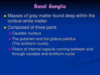
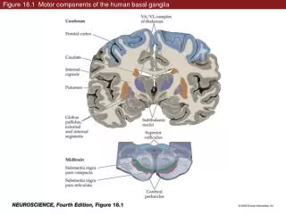
Figure 18.1 Motor components of the human basal ganglia. Figure 18.1 Motor components of the human basal ganglia (Part 1). Figure 18.1 Motor components of the human basal ganglia (Part 2). Figure 18.2 Anatomical organization of the inputs to the basal ganglia.

E N D
Figure 18.1 Motor components of the human basal ganglia (Part 1)
Figure 18.1 Motor components of the human basal ganglia (Part 2)
Figure 18.2 Anatomical organization of the inputs to the basal ganglia
Figure 18.3 Neurons and circuits of the basal ganglia (Part 1)
Figure 18.3 Neurons and circuits of the basal ganglia (Part 2)
Figure 18.4 Regions of the cerebral cortex that project to the corpus striatum
Figure 18.5 Functional organization of intrinsic circuitry and outputs of basal ganglia
Figure 18.5 Functional organization of intrinsic circuitry and outputs of basal ganglia (Part 1)
Figure 18.5 Functional organization of intrinsic circuitry and outputs of the basal ganglia (Part 2)
Figure 18.6 A chain of nerve cells arranged in a disinhibitory circuit
Figure 18.6 A chain of nerve cells arranged in a disinhibitory circuit (Part 1)
Figure 18.6 A chain of nerve cells arranged in a disinhibitory circuit (Part 2)
Figure 18.7 Basal ganglia disinhibition and the generation of saccadic eye movements
Figure 18.7 Basal ganglia disinhibition and the generation of saccadic eye movements (Part 1)
Figure 18.7 Basal ganglia disinhibition and the generation of saccadic eye movements (Part 2)
Figure 18.8 Disinhibition in the direct and indirect pathways through the basal ganglia
Figure 18.8 Disinhibition in the direct and indirect pathways through the basal ganglia (Part 1)
Figure 18.8 Disinhibition in the direct and indirect pathways through the basal ganglia (Part 2)
Figure 18.9 Center-surround functional organization of the direct and indirect pathways
Figure 18.10 Neurological diseases provide insights into function of the basal ganglia
Figure 18.11 Hypo- and hyperkinetic disorders alter the balance of inhibitory signals in the direct and indirect pathways
Figure 18.11 Hypo- and hyperkinetic disorders alter the balance of inhibitory signals in the direct and indirect pathways (Part 1)
Figure 18.11 Hypo- and hyperkinetic disorders alter the balance of inhibitory signals in the direct and indirect pathways (Part 2)
Figure 18.12 Inactivation of tonically active cells of substantia nigra pars reticulata causes saccades
Box 18D Basal Ganglia Loops and Non-Motor Brain Functions (Part 1)
Box 18D Basal Ganglia Loops and Non-Motor Brain Functions (Part 2)