Download
1 / 88
1.08k likes | 1.96k Vues
TREATMENT OF HYDROCEPHALUS (SHUNT PHYSIOLOGY AND PREVENTION OF INFECTION). Hydrocephalous. An excessive accumulation of CSF within the head due to a disturbance of formation, flow or absorption If left untreated the patient may develop increased intra-cranial pressure (ICP)

E N D
TREATMENT OF HYDROCEPHALUS (SHUNT PHYSIOLOGY AND PREVENTION OF INFECTION).
Hydrocephalous • An excessive accumulation of CSF within the head due to a disturbance of formation, flow or absorption • If left untreated the patient may develop increased intra-cranial pressure (ICP) • Result may be brain damage and/or death
Compliance and the Cranium • The brain and skull contain three primary components: • Brain Tissue • Blood • Cerebrospinal fluid • A change in any one of these components results in adjustment to the other two which is called compliance
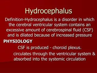
Cerebral Spinal Fluid (CSF) • CSF is produced mainly in the ventricular system by the choroid plexus and is present throughout the central nervous system • CSF has many functions: • Remove waste • Carry nutrition • Regulation of brain function • Neurotransmitter, paracrine and endocrine effects • Cushions the brain
CSF Production & Absorption • CSF is produced at a rate of 20ml per hour and is normally absorbed at the same rate • The brain and spinal cord system contain approximately 150ml of CSF (25ml in the ventricles) • The entire CSF volume is turned over approximately 3 times per day • CSF is transferred from the subarachnoid space into the superior sagittal sinus (venous system) through small granulations called the arachnoid villa
2 Forms of Hydrocephalus • Communicating • Full communication of CSF between ventricles and the subarachnoid space • Non Communicating • CSF cannot flow out of the ventricles due to blockage or malformation
Compliance and Hydrocephalus • As discussed, the skull is a fixed vault with limited volume to hold brain tissue, blood, and CSF • If too much CSF exists, the blood and brain tissue are compressed or squeezed out resulting in a possible neurological deficit
Causes of Hydrocephalus • Acquired hydrocephalus (secondary) • Tumors and cysts • Inflammation (meningitis) • Absorption blockages • Sub Arachnoid Hemorrhage (SAH) • Normal Pressure Hydrocephalus (NPH) • Head injury • Aqueductal Stenosis • Idiopathic hydrocephalus (primary) • iNPH
Symptoms of Hydrocephalus • Usually associated with high intracranial pressure (ICP) • Headaches, nausea, vomiting, sleepiness, irritability, seizures, downward deviation of the eyes, blurred vision, failing mental function, other problems • In infants - expanded head or bulging fontanelles, “sunset sign”.
Pediatric Hydrocephalus • Pediatric Etiologies • Intraventricular hemorrhage (IVH) occurs often in premature babies • Congenital hydrocephalus • Tumor
Adult Hydrocephalus • Adult Etiologies • Normal pressure hydrocephalus (NPH) • Sub arachnoid hemorrhage (SAH) • Post-trauma, aneurysm • Pseudotumor cerebri • Adult onset of congenital hydrocephalus
CT/MRI CRITERIA OF HCP • Hydrostatic hydrocephalus suggested when- • The size of both temporal horns is >2mm in width ; sylvian, interhemispheric fissures and cerebral sulci are not visible. Or • Both temporal horn are >2mm and the ratio FH/ID>0.5
CT/MRI criteria contd.. • Ballooning of frontal horns of lateral ventricles (Mickey mouse ventricles) and third ventricle. • Periventricular low density on CT or periventricular high intensity signal on T2w1 on MRI suggesting transependymal absorption or migration of CSF
CT/MRI criteria contd.. • Used alone- FH/ID <40% - Normal 40-50%- borderline >50% - HCP • Evans Ratio- Ratio of frontal horn to maximal biparietal diameter>30%. • Sagittal MRI may show upward bowing of corpus callosum.
Treating Hydrocephalus • Medical Management. • Spinal Tap. • Surgical Management
Medical treatment • Diuretic therapy- Tried in infants with bloody CSF to see if there is any resumption of normal CSF absorption. • Acetazolamide and furosemide started simultaenously. • To counteract acidosis start alkasol(2meq of K+/ml,no Na +) • S/E- electrolyte imbalance, lethargy, tachypnea, diarrhoea, paraesthesia.
Spinal Taps • HCP after intraventricular hemorrhage may be transient serial taps may temporize until reabsorption resumes but LP can only be performed for communicating HCP. • If reabsorption does not resumes when protein is <100mg/dl then it is unlikely to start as before.
Surgical treatment • Goal- “Optimum neurologic function and good cosmetic result” not “normal sized ventricles. • Options- • Eliminating the cause of obstruction. • Endoscopic methods. • Shunting.
Endoscopic - 3rd Ventriculostomy Indications- Obstructive HCP. Shunt infection(removal of hardware). Patients with subdural hematomas (shunt removed before TV is performed). Slit ventricle syndrome. Contraindication- Communicating HCP.
Endoscopic - 3rd Ventriculostomy contd. • Complications- • Hypothalamic injury. • Transient 3rd and 6th nerve palsies. • Uncontrollable bleeding. • Cardiac arrest. • Traumatic basilar artery aneurysm.
3rd Ventriculostomy contd. • Success rate- overall=56% (range is 60 to 94% for nontumoral aqueductal stenosis). Success rate is lower in infants as they may have under developed sub arachnoid space. • Lower success rate – if preexisting pathology present like- tumor, previous shunt, previous SAH, WBRT, adhesions.
Endoscopic choroid plexus coagulation • First done by Dandy(open) • Indications- • Communicating slowly progressing HCP in infants- 64% cured. • Choroid plexus papilloma/hyperplasia. • Necrotizing enterocolitis. • Intractable shunt failure. • Contraindication- Obstructive HCP.
Endoscopic fenestration • Septostomy – for U/L HCP • Multiloculated HCP. • Aqueductoplasty or aqueductal stenting. • Cysts with secondary HCP- Arachnoid cyst, Cysticercal cysts (3/4 ventricle) • Colloid cyst of third ventricle. • Pineal region tumors- ETV + Biopsy
Types of shunts • VP shunt • VA shunt • Torkildsen shunt- ventricles to cisternal space. • Miscellaneous– Ventriculopleural, gall-bladder, ureter or bladder. • LP shunt • Cyst or subdural shunt
Shunting • Surgical Goal • Re-direct CSF to another area of the body to normalize ICP • Shunt Considerations • Choose the correct operating pressure (fixed pressure valve) • Avoid catheter obstruction • Avoid shunt infection • Avoid other issues • Blood, high protein in CSF • Catheter disconnection
Surgical Procedure • The surgical procedure to place a shunt is relatively short and uncomplicated: • Incision in the scalp • Small burr hole on the skull (6-9mm) • Insertion of the ventricular catheter • Incision in the peritoneal cavity • Tunneling under the skin • Closure
VA Shunt • Repeated lengthening required in a child. • Higher risk of infection and septicemia. • Possible retrograde blood flow into valves. • Shunt embolus. • Vascular complications-thrombophlebitis, pulmonary emboli, PHT
LP Shunt • Do not use in child- laminectomy causes scoliosis, risk of progressive scoliosis • Overshunting. May cause 3rd and 6th CN palsies. • Difficult access to proximal catheter for revision. • Lumbar root irritation. • Leakage of CSF around catheter • Pressure regulation is difficult. • Arachnoiditis.
Torkildsen shunt • Shunts ventricles to cisternal space. • Rarely used. • Effective only in acquired obstructive HCP as children with cong HCP frequently do not develop normal sub arachnoid CSF pathways.
Ventriculopleural shunt • Viable alternative if peritoneum is not available. • Risk of hydrothorax requiring relocation of distal catheter. • Recommended only for >7 yrs of age.
Disadvantages/complications of various shunts • Those that may occur with any shunt- • Obstruction –M.C. , proximal catheter>valve/distal catheter(12-34%) • Disconnection • Infection • Hardware erosion through skin. • Seizures-5.5% in I yr, 1.1 %/yr after 3 Yr.(Higher in frontal catheter.) • Conduit for extraneural mets. • Silicone allergy.
VP shunt complications • Inguinal hernia– if inserted when processus vaginalis is patent. • Requires long catheter to compensate for child growth. • Peritoneal end obstruction-more with distal slit valves, by peritoneal pseudocyst, Peritoneal adhesions may decrease absorptive surface, catheter malpositioning.
VP shunt complications contd.. • Peritonitis • Hydrocele • CSF ascites • Tip migration –Into scrotum, viscus perforation, through diaphragm. • Intestinal obstruction. • Volvulus • Intestinal strangulation- shunt removed forcibly. • Overshunting.
Shunt Complications -INFECTIONS • Incidence -1.5 to 38%(International society of pediatric neurosurgeons cooperative study 1994- 6.5%) • Mortality, morbidity, costs. • Time to infection- 92% infections occur within three months.
Shunt Complications –INFECTIONS contd.. • Risk factors- • Age – most important. (Different skin flora, less immunity and IgG). • Reason for shunt placement. • Type of shunt. • Educational level of surgeon. • Presence of spinal dysraphism.(50% children with MMC who were shunted within 1 wk developed shunt infection.)
Shunt Infections contd.. • Presentation- • Headache,lethargy,nausea and vomiting. • Infants- irritability, severe- apnea and bradycardia. • Fever, gait disturbance, seizures, visual disturbances, upgaze palsy, papilledema, abdominal pain-swelling. • E.coli-severe abdominal pain & septicemia, Staph- indolent , erythema along tract.
Shunt Infections -Evaluation and diagnosis • History and examination (D/D specially in children- URI, gastroenteritis, UTI, Appendicitis) • Imaging- • X ray(shows disconnection of the system.) • USG – Cranium (ependymal enhancement), abdomen. • CT scan • Shunt tap- CSF and manometry
Shunt tap • Indication- • Diagnosing infection/cytology/remove blood/check function/inject medication. 2. Steps- • Insert a 25 gauge butterfly canula and look for flow. Measure pressure. • Measure pressure with distal occlude pressed. • If no spontaneous flow , try to aspirate CSF with a syringe. • Send CSF sample • Connect with manometer. • Repeat measurement after injecting 3-5 ml of saline.
Shunt Infections -Prevention • Sterile surgical technique. • Haines and Walters have found 50% reduction in infections with use of prophylactic antibiotics. • Antibiotic impregnated shunt tubing. • Use of antibiotics before dental procedures, one piece system, biannual screening, hypothermia during surgery.
Treatment outcomes Frame & McLaurin –J neurosurgery.
Shunt Complications- Overshunting 10-12%,VP shunt>VA SHUNT(SIPHONING)
Undershunting • Shunt malfunction rate 17% in I yr. • Cause- blockage (choroid plexus/proteinaceous material/blood); Disconnection. • Symptoms-H/A,N/V, diplopia, lethargy, ataxia, infants, seizures. • Signs-upward gaze palsy, 6thCN palsy, field cut, papilledema.
Shunt Complications (continued) • DisconnectionA part of the system becomes disconnected. The connections between catheters, valves or accessories are damaged. Sometimes due to growth of the patient. • Bowel PerforationThe distal/peritoneal catheter perforates the bowel. Must be revised.
Physiology of shunt devices • History- hippocrates probably did first ventricular puncture. Nulsen and Spitz did the first ventriculojugular Shunt. • Hydrodynamics- • Flow= P/R • R=8nL/^R4 • P=IVP+PGH-OPV-DCP.