Download
1 / 69
720 likes | 1.34k Vues
Histology and Embryology. General Histology. Cells Smallest structures, varies in size, shape and surface. Cells posses properties that permit: Excitability – nerve cells conduct an impulse Synthesis – aiding in the bodies function, such as glands

E N D
Histology and Embryology www.dentalelle.com
General Histology • Cells • Smallest structures, varies in size, shape and surface. • Cells posses properties that permit: • Excitability – nerve cells conduct an impulse • Synthesis – aiding in the bodies function, such as glands • Membrane transport – nutrients are transported • Reproduction – union of sperm and ovum can lead to the formation of an offspring
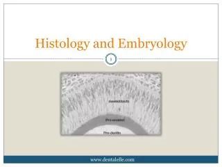
Cell Junctions Desmosomes – cell-to-cell attachments; between ameloblasts and cells of stratified squamous epithelium that lines the oral cavity. Tight junctions – cells attach to each other by fusion of their cell membranes; adjacent odontoblasts form tight junctions that prevent substances in the pulp from passing into the dentin. Gap junctions – channel that runs between cells for communication of cell electrical impulses and passage for molecules; present amount some odontoblasts, allowing to coordinate their activity. Hemidesmosome – attachment of a cell to a noncellular surface; basal layer cells of stratified squamous epithelium attach to the basement membrane by hemidesmosomes; present in epithelial attachment of the tooth
The Cell • Cells are surrounded by a cell membrane that separates them from the outside environment. • 1. cytoplasm • 2. organelles • 3. inclusions • 4. nucleus • Specialization • A) differentiation – cells that recognize one another will group together • B) organization of chemicals – chemicals appear earlier in the embryo. Endocrine substances are produced by one type of cell and can affect other types of cells • C) Cells tissues organs organ systems
Cell Membrane • Cell membrane • A) Called a plasma membrane or plasmalemma; selectively permeable because it controls passage of materials in and out of the cell. Uses active transport, passive transport, or facilitated diffusion. • Lipids and proteins are the major components (3:2 ratio of proteins) • Structure is trilaminar, with bipolar membrane and a central core of lipids between two layers of protein • Diffusion of small lipid-insoluble substances
Fluid Mosaic Model • Shown on the previous slide • Trilaminar structure composted of two facing layers of lipid molecules, into which larger globular proteins are inserted. • Lipid bilayers consist mainly of phospholipid molecules; hydrophilic ends face the outer and inner surfaces of the cell; the hydrophobic ends attract and face each other. • Globular proteins are integral proteins and peripheral proteins. Integral extend through the full width of the cell membrane and protrude and may have carbohydrate units attached to them. Peripheral are linked or attached to the cell membrane surface.
Cytoplasm • Translucent, aqueous, homogeneous gel enclosed in the cell by the cell membrane. • The cytoplasm has three major elements; the cytosol, organelles and inclusions. • All metabolic activities of the cell occur in the cytoplasm which include: • Assimilation (digestion) • Synthesis of substances such as proteins, proteoglycans, and glycoproteins • A transport medium in which all nutrients and metabolites are carried from one organelle to another • Presence of enzymes and electrolytes where specific metabolic reactions take place (glycolysis)
Nucleus In cell biology, the nucleus is a membrane-enclosed organelle found in eukaryotic cells. It contains most of the cell's genetic material, organized as multiple long linear DNA molecules in complex with a large variety of proteins, such as histones, to form chromosomes. The genes within these chromosomes are the cell's nuclear genome. The function of the nucleus is to maintain the integrity of these genes and to control the activities of the cell by regulating gene expression — the nucleus is, therefore, the control center of the cell. The main structures making up the nucleus are the nuclear membrane, a double membrane that encloses the entire organelle and isolates its contents from the cellular cytoplasm, and the nucleoskeleton (which includes nuclear lamina), a mesh work within the nucleus that adds mechanical support, much like the cytoskeleton, which supports the cell as a whole.
Nucleus Nuclear pores are required that regulate Nuclear transport of molecules across the envelope. The pores cross both nuclear membranes, providing a channel through which larger molecules must be actively transported by carrier proteins while allowing free movement of small molecules and ions. The interior of the nucleus does not contain any membrane-bound sub compartments. The best-known of these is the nucleolus, which is mainly involved in the assembly of ribosomes. After being produced in the nucleolus, ribosomes are exported to the cytoplasm where they translate mRNA.
Synthesis Activities • Three types of RNA are necessary for protein synthesis: • Messenger RNA (mRNA) – copies of short segments of deoxyribonucleic acid (DNA) • Contains all genetic information of proteins • Must pass through the ribosomes attached to the endoplasmic reticulum • As it passes through the ribosomes, transfer RNA (tRNA) adds the exact amino acid to the newly forming proteins • Protein synthesis can also occur on polyribosomes floating freely in the cytoplasm; proteins synthesized on the ribosomes attached to the ER are transported out of the cell
Inclusions Inclusions are nonliving metabolic by-products found in the cytoplasm. May appear as lipid droplets, carbohydrate accumulations, or engulfed foreign substances
Lysosomes • Intracellular digestion is carried out by organelles called lysosomes. There are several contexts in which cells need to carry out digestion. • Include the recycling of cellular organelles and the breakdown of viruses and other cellular invaders. Single-celled organisms use lysosomes to digest their food as they have no process for extracellular digestion. The pH within a lysosome is very acidic and the enzymes within work most effectively in this environment. • The components of a lysosome have evolved specific conformations that make them resistant to break down by the enzymes within the lysosome. • During phagocytosis, lysosomes fuse with engulfed substances to form a secondary vesicle; the vesicle may then remain in the cell as a residual body or discharged outside the cell
Golgi Complex The Golgi apparatus receives protein and/or lipid-filled vesicles that bud from the ER. The Golgi apparatus contains enzymes that modify proteins and lipids. For example, it can add a chain of sugars to proteins and lipids, thereby making them glycoproteins and glycolipids, which are molecules found in the plasma membrane. The vesicles that leave the Golgi apparatus move to other parts of the cell. Some vesicles proceed to the plasma membrane where they discharge their contents. Because this is secretion, note that the Golgi apparatus is involved in processing, packaging, and secretion. Other vesicles that leave the Golgi apparatus are lysosomes. The Golgi complex is the storage site for newly synthesized proteins and of course packaging and transporting many cell products. Also produces large carbohydrate molecules and lysosomes.
Mitochondria Although the size and shape of mitochondria (sing., mitochondrion) can vary, all are bounded by a double membrane. The inner membrane is folded to form little shelves called cristae, which project into the matrix, an inner space filled with a gel-like fluid. Mitochondria are the site of ATP (adenosine triphosphate) production involving complex metabolic pathways. As you know, ATP molecules are the common carrier of energy in cells. A shorthand way to indicate the chemical transformation that involves mitochondria.
Mitochondria Continued Mitochondria are often called the powerhouse of the cell: Just as a powerhouse burns fuel to produce electricity, the mitochondria convert the chemical energy of carbohydrate molecules into the chemical energy of ATP molecules. Inthe process, mitochondria use up oxygen and give off carbondioxide and water. The oxygen you breathe in enters cells and then mitochondria; the carbon dioxide you breathe out is released by mitochondria. Because oxygen is used up and carbon dioxide is released, we say that mitochondria carry on cellular respiration.
Endoplasmic Reticulum The endoplasmic reticulum (ER), a complicated system of membranous channels and flattened vesicles, is physically continuous with the outer membrane of the nuclear envelope. Rough ER is studded with ribosomes on the side of the membrane that faces the cytoplasm. Here proteins are synthesized and enter the ER interior where processing and modification begin. Some of these proteins are incorporated into membrane, and some are for export. Smooth ER, which is continuous with rough ER, does not have attached ribosomes. Smooth ER synthesizes the phospholipids that occur in membranes and has various other functions, depending on the particular cell. In the testes, it produces testosterone, and in the liver it helps detoxify drugs. Regardless of any specialized function, ER also forms vesicles in which large molecules are transported to other parts of the cell. Often these vesicles are on their way to the plasma membrane or the Golgi apparatus.
Filaments and Tubules Associated with contractility in cells – thread-like structures about 7-10nm thick Microfilaments act as a support system for the cell cytoskeleton Bundles of microfilaments form tonofibrils and become part of the attachment apparatus between cells (desmosomes)
Microtubules Delicate tubes, 20-27 nm wide, found in cells that are undergoing mitosis and alterations in cell shape They have an internal support function, especially in long cellular processes such as neurites or odontoblastic processes They have the capacity to direct intracellular transport through the cytoplasm
Centrioles Cylindrical structures composed of microtubule like components Centrioles function in cell replication and the formation of cellular extensions
Internal Environment and Homeostasis • Extracellular fluid • Circulates outside and between cells • Intracellular fluid • Fluid located inside the cells of the body • Homeostasis • The delicate balance maintained between the two fluid compositions
Transport through the Cell Membrane Diffusion - is the random movement of simple atoms or molecules from area of higher concentration to an area of lower concentration until they are equally distributed. To illustrate diffusion, imagine putting a tablet of dye into water . The water eventually takes on the color of the dye as the dye molecules diffuse. The chemical and physical properties of the plasma membrane allow only a few types of molecules to enter and exit a cell by simple diffusion. Lipid-soluble molecules such as alcohols can diffuse through the membrane because lipids are the membrane’s main structural components. Gases can also diffuse through the lipid bilayer; this is the mechanism by which oxygen enters cells and carbon dioxide exits cells. For example, consider the movement of oxygen from the lungs to the bloodstream.
Transport Continued When you inhale, oxygen fills the tiny air sacs, or alveoli, within your lungs. Neighboring lung capillaries contain red blood cells with a very low oxygen concentration. Oxygen diffuses from the area of highest concentration to the area of lowest concentration: first through alveolar cells, then lung capillary cells, and finally into the red blood cells. When atoms or molecules diffuse from areas of higher to lower concentration across plasma membranes, no cellular energy is involved. Instead, kinetic or thermal energy of matter is the energy source for diffusion.
Osmosis Osmosis is the diffusion of water across a plasma membrane. Osmosis occurs whenever an unequal concentration of water exists on either side of a selectively permeable membrane. (Recall that a selectively permeable membrane allows water to pass freely, but not most dissolved substances.) In a solution, water is more concentrated when it contains fewer dissolved substances, or solutes, (and thus is closest to pure water). Water is less concentrated as solute concentration increases. Osmotic pressure is the force exerted on a selectively permeable membrane because water has moved from the area of higher water concentration to the area of lower water concentration(higher concentration of solute).
Osmosis Continued Tonicity is the degree to which a solution’s concentration of solute-versus-water causes water to move into or out of cells. Normally, body fluids are isotonic to cells that is, there is an equal concentration of solutes (dissolved substances) and solvent (water) on both sides of the plasma membrane, and cells maintain their usual size and shape. Medically administered intravenous solutions usually have this tonicity. Body fluids which are not isotonic to body cells are the result of dehydration or water intoxication. Solutions (solute plus solvent) that cause cells to swell or even to burst due to an intake of water are said to be hypotonicsolutions. If red blood cells are placed in a hypotonic solution, which has a higher concentration of water (lower concentration of solute) than do the cells, water enters the cells and they swell.
Continued The term lysis refers to disrupted cells: hemolysis, then, is disrupted red blood cells. Solutions that cause cells to shrink or to shrivel due to a loss of water are said to be hypertonic solutions. If red blood cells are placed in a hypertonic solution, which has a lower concentration of water (higher concentration of solute) than do the cells, water leaves the cells and they shrink.
Active Transport • Process used by the cell when large quantities of a substance are needed inside the cell and only a small amount of the substance is present in the extracellular fluid. • Pumps the substance against its concentration gradient. • ATP • Sodium pump; important for the transmission of nerve impulses • Almost all monosaccharides are actively transported into the body. • Phagocytosis – movement of a solid particle into the cell • Pinocytosis – movement of fluid into a cell, the cell invaginates around fluid
Cell Replication - Mitosis Interphase: During interphase, the cell carries on its regular activities, and it also gets ready to divide if it is going to complete the cell cycle. For these cells, interphase has three stages, called G1 phase, S phase, and G2 phase. G1 Phase - it is best to think of G as standing for “growth.” Protein synthesis is very much a part of these growth phases. During G1, a cell doubles its organelles (such as mitochondria and ribosomes) and accumulates materials that will be used for DNA synthesis.
Continued S Phase Following G1, the cell enters the S (for “synthesis”) phase. During the S phase, DNA replication occurs. At the beginning of the S phase, each chromosome is composed of one DNA double helix, which is equal to a chromatid. At the end of this phase, each chromosome has two identical DNA double helix molecules, and therefore is composed of two sister chromatids. Another way of expressing these events is to say that DNA replication has resulted in duplicated chromosomes. G2 Phase During this phase, the cell synthesizes proteins that will assist cell division, such as the protein found in microtubules. The role of microtubules in cell division is described later in this section.
Prophase Several events occur during prophase that visibly indicate the cell is about to divide. The two pairs of centrioles outside the nucleus begin moving away from each other toward opposite ends of the nucleus. Spindle fibers appear between the separating centriole pairs, the nuclear envelope begins to fragment, and the nucleolus begins to disappear. The chromosomes are now fully visible. Spindle fibers attach to the centromeres as the chromosomes continue to shorten and thicken. During prophase, chromosomes are randomly placed in the nucleus.
Prophase Continued At the end of prophase, a cell has a fully formed spindle. A spindle has poles, asters, and fibers. The asters are arrays of short microtubules that radiate from the poles and the fibers are bundles of microtubules that stretch between the poles. (A spindle resembles a lopsided bicycle wheel; the asters are the “spokes.”) Centrioles are located in centrosomes, at opposite poles of the cell. Centrosomes are believed to organize the spindle.
Metaphase During metaphase, the nuclear envelope is fragmented, and the spindle occupies the region formerly occupied by the nucleus. The paired chromosomes are now at the equator (center) of the spindle. Metaphase is characterized by a fully formed spindle, and the chromosomes, each with two sister chromatids, are aligned at the equator
Anaphase At the start of anaphase, the sister chromatids separate. Once separated, the chromatids are called chromosomes. Separation of the sister chromatids ensures that each cell receives a copy of each type of chromosome and thereby has a full complement of genes. During anaphase, the daughter chromosomes move to the poles of the spindle. Anaphase is characterized by the movement of chromosomes toward each pole and thus, to opposite sides of the cell.
Telophase A nuclear membrane forms around each set of chromosomes Centrioles replicate in each cell
Dental Tissues Organelles play an important role in providing energy (mitochondria) which helps in calcifying dental tissues Cell organelles help maintain tissues after the initial formation by the cell; fibroblasts contain increased numbers of cell organelles; these additional organelles aid fibroblasts in their synthesizing and secretory functions
Basic Tissues • Individual cells multiply and differentiate to perform specialized functions; groups of cells with similar characteristics and functions come together and form tissues. • Tissue components • Cells • Intercellular substance – product of living cells, passageway • Tissue Fluid – blood plasma that transports
Tissues in the Human Body 1. epithelial tissues (covering/lining); 2. connective tissues (support); 3. muscle tissues (movement); 4. nervous tissues (control).
Epithelial Tissue Many epithelial tissues are classified according to their shape and the number of layers they possess: Some terms used to describe epithelia include: a. simple = single layer of cells; b. stratified = many layers of cells; c. squamous = flattened cells; d. cuboidal = square-shaped cells; e. columnar = elongated cells (i.e. taller than wide);
Types of Simple Epithelium • Simple squamous epithelium: • a single layer of flattened cells; • generally allows for easy passage (diffusion) of substances; • Locations: 1. lining air sacs of lungs, 2. lining capillaries, 3. lining body cavities, 4. covering ventral organs;
Simple Cuboidal Epithelium • Simple cuboidal epithelium: • a single layer of square-shaped cells with large centrally located nuclei; • Functions: 1. secretion 2. absorption; • Locations: 1. lining kidney tubules, 2. lining ducts of glands, 3. covering surface of ovary;
Simple Columnar Epithelium • Asingle layer of elongated cells with basally located nuclei (near basement membrane); 1. protection, 2. absorption, 3. secretion; 1. lining small intestine, 2. lining uterus; • Free Surface Modifications: 1. microvilli (increase surface area) 2. goblet cells (secrete protective mucus);
Pseudostratified Columnar Epithelium • Pseudostratified columnar epithelium:a single layer of elongated cells with scattered nuclei (i.e. look stratified but are not); all cells touch the basement membrane • Functions: 1. secretion, 2. protection; Locations: 1. lining trachea, 2. lining fallopian tube; • Free surface modifications: 1. cilia (trap debris and aid in passage of mucus up and out of airway); 2. goblet cells (produce mucus which coats cilia and helps trap debris).
Stratified Epithelium Stratified squamous epithelium: many layers of flattened cells; Function = protection; Locations: Non-keratinized: 1. lining mouth, 2. lining throat, Keratinized – epidermis of the skin
Stratified Cuboidal Epithelium • Stratified Cuboidal epithelium:2-3 layers of cuboidal cells. • Locations 1. mammary glands 2. sweat glands 3. salivary glands 4. pancreas