Download
1 / 27
380 likes | 846 Vues
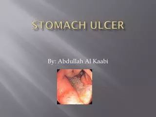
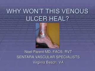
WHY WON’T THIS VENOUS ULCER HEAL?. Noel Parent MD, FACS, RVT SENTARA VASCULAR SPECIALISTS Virginia Beach, VA. Variety of leg ulcer etiology. Decubitis (pressure) at points of contact Ischemic (arterial) malperfusion Infectious Foreign body Rheumatologic or autoimmune Venous

E N D
WHY WON’T THIS VENOUS ULCER HEAL? Noel Parent MD, FACS, RVT SENTARA VASCULAR SPECIALISTS Virginia Beach, VA
Variety of leg ulcer etiology • Decubitis (pressure) • at points of contact • Ischemic (arterial) • malperfusion • Infectious • Foreign body • Rheumatologic or autoimmune • Venous • Venous hypertension
Venous ulcer characteristics • Ulcer location is medial lower calf just above the ankle • Hemosiderin deposition • Lipodermatosclerosis • Edema • Size variable • Varicose veins +/-
It all begins with loss of venous valve function (venous reflux)
Elevated pressure within the veins of the calf skin is the pathophysiology
…did not heal in the past: INADEQUATE DIAGNOSTIC IMAGING IMPRECISE INVASIVE TREATMENTS …why they may heal now: DUPLEX ULTRASOUND IMAGING FOR VEIN MAPPING EVLT IS PRECISE AND MINIMALLY INVASIVE Why vein ulcers…
Duplex ultrasound perforator vein imaging to access for ablation
Very dirty ankle venous ulcer in an elderly female. Complex PVL showing: chronic thrombus in GSV. Note associated SSV reflux and two incompetent calf perforator veins. Case example #1
PVL confirms successful ablation of GSV, SSA and perforators. 6 weeks post endovenous laser treatment: ulcer healing for the FIRST TIME IN A YEAR! Case example #1, cont’d
Pre treatment Post treatment Case example #2
Summary • Leg ulcers are commonly due to elevated venous pressure in skin from valve weakness resulting in reflux. • Results from traditional therapies of Unna boots, stockings, and open surgery are disappointing. (Costly, morbid, disabling) • A contemporary treatment consisting of duplex scan diagnosis of perforating veins, then treatment by EVLT, may become the new standard of care.