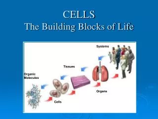
Basic Building Blocks: Proteins
Basic Building Blocks: Proteins. Largest variety of biomolecules Most of the weight of cells, aside from water Basic unit is amino acid Form of amino acid Simplest is glycine with R = H All others are asymmetric two stereoisomers L & D with mainly L naturally occuring.


Basic Building Blocks: Proteins
E N D
Presentation Transcript
Basic Building Blocks: Proteins Largest variety of biomolecules Most of the weight of cells, aside from water Basic unit is amino acid Form of amino acid Simplest is glycine with R = H All others are asymmetric two stereoisomers L & D with mainly L naturally occuring
Human Genome Project Facts • Human DNA codes about 30,000 genes (vs. fruit flies:13,500 and C. elegans: 19,000) • These genes represent only ~ 1% of DNA – lots of coding for control & transcription factors • Average human protein has ~450 amino acids • One of the largest proteins is titin (27,000 amino acids in a single chain)
Protein Functions • Motion & locomotion of cells/organism (contractile proteins) • Catalysis of all biochemical reactions (enzymes) • Structure of cells and extracellular matrix (e.g. collagens) • Receptors for hormones/ signaling molecules • Transcription factors • Etc.
Example Protein (H-2K) - Structure • This antigen displays many features of proteins • Two polypeptide chains • Longer heavy chain has 5 domains – 3 extracellular, one transmembrane, and one cytoplasmic – it is called an integral membrane protein • Smaller polypeptide chain is attached to heavy chain by H bonds (no covalent bonds) – it is a peripheral membrane protein • The dark bars are disulfide bridges (S-S) • Two short branched sugars are on the left making this a glycoprotein (sugar + protein compex) The view seen here does not show its real 3D arrangement Look in PDB
Types of amino acids • Classify aa by various criteria – each has 3 letter or 1 letter code • 3 have ring-structures – important in fluorescence • All are ampholytes (+/- charge depending on pH)
Digression: pH ideas • pH = -log[H+] • Neutrality when [H+]=[OH-]=10-7 M • Higher pH – basic; lower – acidic • Simple idea: H2O OH- + H+ • Dissociation constant K where G = free energy per mole of bond formation; with [H2O] = 55 M ~ constant So K’ = [H+][OH-] = 10-14 and pK = -log K in general
pH and pK • Each charged group has a pK • For proteins, e.g., • COOH COO- + H+ pK 2.34 • NH3+ NH2 + H+ pK 9.69 • R group dissociation also If pH > pK more basic form If pH < pK more acidic form Different forms predominate at different pH - polyelectrolyte
Example: Titration of alanine • Different forms at different pH • Alanine has R = CH3 • pI = isoelectric point = pH at which neutral
Peptide bond • Amino acids link together to form a continuous linear chain = backbone of protein
Primary Structure • With even only 10 a.a. long – number of possible polypeptides (decamers) = 2010 = 1010x210 ~ 1013 • Amino acid composition – not sequence – can be automatically determined by aa analyzer to give % composition • General features of 1o structure: • Most polypeptide chains are 100 – 500 aa; smallest 25 – 100, largest 3000 • Some proteins have more than 1 chain – held together by weaker non-covalent bonds • Protein data bank – on-line
Facts about 1o structure • Wide variation in composition • Certain aa are fairly rare (methionine, Tryptophan) • Ala, Leu very common • Many proteins contain other molecules, including carbohydrates, metal ions (Ca, Fe, Zn, Cu)
Secondary Structure (2o) of Proteins • Backbone of protein chain has series of rotatable bonds. Two angles describe possible rotations of each peptide • Rotations about these bonds lead to certain allowed structures – or stable conformations
Ramachandran Diagram • A number of helices and b sheets are possible
a-helix + b-sheet • Pairs of chains lying • side by side • - Stabilized by H bonds - Right-handed - 3.6 aa per turn - R groups outside - H bond between -NH and –C=O 4 aa apart pointing along axis
More a-helix, b sheet, triple helix Collagen triple helix All proteins consist of regions of 2nd structure w/ random coil connections
Prediction of structure • Based on knowing aa sequence, we are able to predict a-helix, b-sheet regions • For example: residues 1-36 in histone have 12 + charges – able to bind to neg. charges on d-s DNA • For example: glycophorin from human RB cells spans membrane from 73 – 95 non-polar region
Protein Folding Problem • Big Question is: If you know the primary sequence of aa can you predict the 3-D structure of a protein? [Protein-folding problem – one of challenges] • Can occur spontaneously – involves basic electrical interactions that we’ll study soon • Co-valent bonds along backbone • H-bonds – weaker, directional • Van der Waals – non-specific attractive • Hydrophobic/ hydrophilic – entropy driven forces
Tertiary Structure (3o) • All proteins consist of 2o structure regions connected by random coil • Human ICE-protease Interleukin-1ß-converting enzyme
Protein Domains • Tertiary structure of proteins is built up from domains • Each domain has a separate function to perform – for example: • Binding a small ligand • Spanning the plasma membrane • Containing a catalytic site • DNA binding (transcription factors) • Providing a binding surface for another protein • Often each domain is encoded by a separate exon in the gene encoding that protein – this correspondence is most likely to occur in recently-evolved proteins (exon shuffling idea to generate new proteins using established domains – like Lego pieces)
Fibrous Proteins • Two major classes of proteins based on 3o Structure • Fibrous – fiber-like, includes • Keratins – in hair, horns, feathers, wool • Actin – muscle thin filaments, cells • Collagen – connective tissue Often these are polymers made up from monomer subunits and form all a helices and/or all b sheets (e.g. silk) Actin filament made from monomers
Globular Proteins • Second class is globular– most enzymes, hormones, transport proteins – folded up structure
General Properties of 3o Structure • Lowest energy states are most stable 3o structures • Charged residues are on surface or exposed clefts • Non-polar (hydrophobic) residues are internal • Nearly all possible H-bonds form
Quaternary (4o) Structure • Multiple sub-units bound together non-covalently • Canonical example is hemoglobin:
Cooperative Binding by Hemoglobin • Fe in the heme group binds oxygen – separately, each of 4 hemes binds O2 as in myoglobin – 4 together bind O2 cooperatively – Allosteric conformational change Sigmoidal binding curve indicates cooperativity
Packing Density of Proteins • How filled is volume of protein? • Quantitative measure = packing density = • For continuous solid PD = 1 • For close packed spheres PD = 0.74 • For close packed cylinders PD = 0.91 • For ribonuclease S, PD = 0.75
Two Other Classes of Biomolecules- Polysaccharides + Lipids • Polysaccharides (carbohydrates) • Monosaccharide – eg glucose • Disaccharide – eg lactose • Polymers of sugars – M ~ 104 – 107 Da lactose +
Polysaccharides - con’t • Glucose can polymerize into 3 types of polymers • Starch- polymers of glucose – metabolic • Glycogen- ditto, but with more shorter branching –also metabolic- stores glucose • Cellulose – most prevalent biomolecule - structural
Lipids • Very diverse family – all insoluble in water/ rich in hydrocarbons • Includes fatty acids, steroids, phosphoglycerides/phospholipids in membranes • Polar head group = fatty acid tail with 12 – 24 C’s in tail cholesterol
micelle bilayer Vesicle (unilamellar)