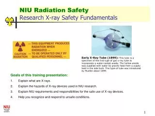
ANALYTICAL X-RAY SAFETY User Training
Centre for Environmental Health, Safety and Security Management. ANALYTICAL X-RAY SAFETY User Training. Analytical X-ray Safety Training – User Training TRAINING OUTLINE. History Sources/uses of X-rays Legislation Biological & Health Effects X-ray safety in the lab Exposure SOPS

ANALYTICAL X-RAY SAFETY User Training
E N D
Presentation Transcript
Centre for Environmental Health, Safety and Security Management ANALYTICAL X-RAY SAFETYUser Training
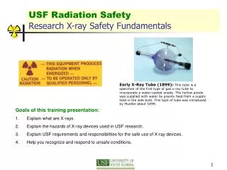
Analytical X-ray Safety Training – User Training TRAINING OUTLINE • History • Sources/uses of X-rays • Legislation • Biological & Health Effects • X-ray safety in the lab • Exposure • SOPS • Security • Emergencies • Summary • References • Quiz RYERSON UNIVERSITY
IN THE BEGINNING… RYERSON UNIVERSITY
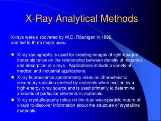
Wilhelm Roentgen (1845-1923)Discovers X-Rays • German physicist discover X-rays November 8, 1895 • Studying cathode ray tubes, noticed that the fluorescence occurred even when outside light was shielded by black paper wrapped around cathode ray tube • Discovery instantly revolutionized physics and medicine; lead to another field of research of radioactivity • 1901 Nobel prize in physics awarded for his discovery RYERSON UNIVERSITY
Henri Becquerel (1852-1908)Discovers Radioactivity • French physicist discovers radioactivity March 1, 1896 • Believed sun’s rays were absorbed by uranium then emitted as x-rays • Due to overcast skies, returned uranium rocks to storage drawer on top of photographic plates • Developed plates showed clear and strong images i.e., spontaneous emission of radiation by anatural material • Shares Nobel Prize with Curies 1903 RYERSON UNIVERSITY
Marie Sklodowska Curie (1867-1934)Double Nobel prize winner in physics & chemistry • Contributes to W W 1 French war effort by making public pleas for fund to equip ambulances with radiology equipment • Elected by Red Cross to be official head of Radiological Service • Devised courses in radiology and taught doctors new techniques to locate foreign objects in the human body RYERSON UNIVERSITY
Nobel Prizes forResearch using X-Rays 1901 (Physics) W.C. Roentgen discovery of X-Rays 1914 (Physics) M.von Laue x-ray diffraction from crystals 1924 (Physics) W. H. Bragg & W. L. Bragg crystal structure from x-ray diffraction 1917(Physics) C. G. Barkla characteristic radiation of elements 1924 (Physics) K. M. G. Siegbahn x-ray diffraction 1927(Physics) A. H. Compton scattering of x-rays by electrons RYERSON UNIVERSITY
Nobel Prizes forResearch with X-Rays 1936 (Chemistry) P. Debyediffraction of x-rays & electrons in gases 1962 (Chemistry) M. Perutz & J. Kendrewstructure of hemoglobin 1979 (Medicine) A. McLeod Cormack & G. Newbold Hounsfield computed axial tomography 1981 (Physics) K. M. Siegbahnhigh resolution electron spectroscopy 1985 (Chemistry) H. Hauptman & J. Karle direct methods to determine x-ray structures 1988 (Chemistry) J. Deisenhofer, R. Huber & H. Michel structures of proteins crucial to photosynthesis RYERSON UNIVERSITY
Wimshurt Static Machine(circa 1890s) • High voltage for x-ray tubes was provided by a static machine or an induction coil • Static machines were cheap and simple setup but could not provide as high a current as induction coils • Some machines used more than 12 discs up to 3 ft in diameter RYERSON UNIVERSITY
Induction Coil (circa 1900) • Until around 1910, the high voltages (104 volts required for x-ray tube operation provided by induction coils • Operated off DC current provided by battery cells • Eventually replaced by transformers RYERSON UNIVERSITY
X-ray Timer (French)(circa 1900-1920) • Timer permitted x-ray tube to be operated for up to 11 sec • Physician turned the dial to chosen exposure, pressed button on top of clock • Connected high voltage line (from induction coil or static machine) to x-ray tube • 2 electrical terminals located below clock face RYERSON UNIVERSITY
commonly seen in shoe stores in the 1930-1950s vertical cabinet with an opening at the bottom into which the feet were placed. image of the bones of the feet and the outline of the shoe could be seen through each of the three viewing ports on the top of the cabinet Shoe-Fitting Fluoroscope (ca. 1930-1940) RYERSON UNIVERSITY
Elephants???? Globe and Mail article February 3, 2004 RYERSON UNIVERSITY
Continue on to:Sources and Uses Return to Main Menu RYERSON UNIVERSITY