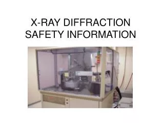
X-RAY DIFFRACTION SAFETY INFORMATION
X-RAY DIFFRACTION SAFETY INFORMATION. Restricted Item List. Any proposed purchase or acquisition, and installation of x-ray equipment must be reviewed and approved in advance by the Tufts Radiation Safety/EHS. Radiation symbols inform persons of radiation producing

X-RAY DIFFRACTION SAFETY INFORMATION
E N D
Presentation Transcript
Restricted Item List Any proposed purchase or acquisition, and installation of x-ray equipment must be reviewed and approved in advance by the Tufts Radiation Safety/EHS. Radiation symbols inform persons of radiation producing equipment or RAM use.
Caution – XRD can be hazardous Detailed instructions on the operation, hazards, and radiation safety features of a specific analytical unit must be provided by the owner of the equipment. Before starting to work on an analytical unit, make sure you receive specific instruction on the unit’s safe operation from the Tufts person responsible for the unit. All x-ray users must have attended Tufts radiation safety training, but that is NOT a substitute for unit specific operation and safety training.
Introduction Analytical x-ray devices are important tools in various areas of modern research. X-ray crystallography and x-ray fluorescence spectrometry rely on x-radiation. But, X-ray diffraction equipment [XRD] can be very dangerous, and operators of this equipment must not become complacent or overconfident about the potential danger of the x-ray beam.
X-ray Production When high energy electrons strike an anode in a sealed vacuum, x-rays are generated. Anodes are often made of copper, iron or molybdenum. X-rays are electromagnetic radiation. They have enough energy to cause ionization.
Radiation Units Roentgen R – unit of exposure, in air for photons only. One R equals enough energy to deposit 2.58 x 10 -4 coulombs per kg in dry air. Rad – unit of absorbed dose. Equal to one hundred ergs per gram Rem – unit of dose equivalent. For x-rays, 1 rad = 1 rem Milli – 1/1000th, as in millirem or mRem
Typical X-ray Beam Intensities* Primary beam 400,000 Rem/min, or 2.4 x 10 7 Rem/hr Diffracted beam 80 Rem/hr * For comparison, the annual whole body occupational exposure limit is 5 Rem. Radio- isotope users at Tufts generally receive < 100 mRem/yr.
MA DPH Occupation Exposure Limits Area: Annual dose limit, Rem: Whole body, lens of eye, head, trunk 5 Hands, arms, 50 skin of whole body
ALARA ALARA stands for as low as reasonably achievable. Regulators recognize that it is an individual worker’s responsibility to perform tasks on a daily basis keeping best practices in mind, and striving to keep radiation exposure as low as possible. Worker’s are responsible for knowing all hazards and safety practices that relate to the equipment in use.
Biological Effects of Radiation Effect Dose, Exposure time in Rem primary beam, seconds Erythema 300-600 0.075-0.12 Epilation 350 temporary 0.0525 1200 permanent 0.180 Acute dermatitis 3000-4000 0.45-0.60 Chronic dermatitis thousands of Rem in many small doses over NA many years Skin Cancer small doses over a long ??? period of time
GENERAL RADIATION INFORMATION IONIZING RADIATION CAN CAUSE CHEMICAL CHANGES IN BIOLOGICAL TISSUE. THESE CAN LEAD TO CELL DEATH, CELL TRANSFORMATION, AND DAMAGE WHICH CAN NOT BE REPAIRED.
WARNING Very serious injuries have resulted from the use of XRD equipment. Large doses of radiation have caused painful burns and permanent injuries to workers.
You could be injured without initially knowing it - People are not able to sense radiation. Even very large doses of radiation can not be felt. Just because there was no sensation at the time the dose is received, does not mean you are safe. Serious injury can result for radiation exposure. It is up to the individual x-ray user to ensure that they are trained, follow all precautions, and use all x-ray equipment safely.
Sources of Exposure The primary beam, Leakage of primary beam through cracks in shielding, Penetration of primary beam through shutters, cameras, beam stops, etc., Secondary emission (fluorescence) from a sample or shielding material, Diffracted rays from crystal, Radiation generated by rectifiers in the high voltage power supply of older units.
Sealed Tube/Microfocus Systems:What are the danger areas? 3. Leakage 1. Primary Beam 2. Scattered Radiation
Three regions of high exposure include the primary beam, scattered radiation, and leakage radiation.
1.Primary Beam The critical radiation exposure problem with analytical X-ray equipment is the primary beam. Exposure to the primary beam can cause localized acute exposure. Consequently, the analytical operator must never intentionally place any part of their body in the primary beam. Typically, these beams are relatively “soft” X-rays resulting in maximal energy deposition in epithelial tissues. Erythema or reddening of the skin can occur when skin is acutely exposed to 300-600 R (much less than a second). Radiation burns may occur from longer exposures. 2.Scattered Radiation When the primary beam intersects a material such as a sample or elements of the X-ray unit including the beam stop, some of the radiation is scattered out of the primary beam. While these radiation fields are considerable less intense than the primary beam, they still represent a potential hazard. Scattered radiation fields can be measured by the analytical operators with a survey meter. 3.Leakage Some radiation may leak around the tube housing structure. The source housing construction must be such that when all the shutters are closed, the leakage radiation must not exceed that of radiation limits for the general public.
Instruments which are calibrated for radiation that uniformly exposes the active area of the detector will give incorrect low readings when exposed to a beam having a smaller area. To determine the true reading, the measured reading must be multiplied by f, where f = area of detector/area of beam Check instrument batteries Have audio “on” Begin on “fast” or “F” setting Precise measurements may be taken using “slow” or “S” setting, as the needle will not “bounce” as much Scales will vary with each instrument model Begin on X1 scale Radiation monitoring instruments
Safety Basics • Time – minimizing time around a radiation source will reduce total exposure • Distance – maximize distance from a radiation source to reduce total exposure See “Inverse Square Law” • Shielding – material used to attenuate radiation and reduce occupational exposure. For x-rays, shielding is most often lead.
Inverse Square Law Radiation exposure varies inversely as the square of the distance from the source E ∞ 1 / d 2
DETAILED INSTRUCTION ON THE SAFE USE OF XRD MUST BE PROVIDED BY THE SUPERVISING PI AND/OR HIS/HER DESIGNEE. THIS INSTRUCTION MUST INCLUDE DEMONSTRATION OF ALL SAFETY FEATURES OF ALL SPECIFIC EQUIPMENT TO BE USED. BEFORE BEGINNING USE OF ANY XRAY EQUIPMENT, BE SURE YOU UNDERSTAND ALL OF THE TRAINING. IF PROBLEMS OR QUESTIONS ARISE, STOP WORK AND CONSULT YOUR SUPERVISOR AND THE EQUIPMENT OWNER/SUPERVISOR.
Characteristics of XRD Beams Both primary and diffracted beams are generally small and well collimated.
Wavelengths used in crystallography are often in the range of 0.6 to 2.5 A. The 1.54 A wavelength corresponds to CuKa radiation.
Safety Devices and Features All units require a clear, visible warning light what illuminates only when the unit is producing X-rays. Shutter status shall be indicated clearly. Shutters must not be able to open without a collimator or coupling device in place. Safety interlocks shall not be bypassed or defeated. Unused ports shall be secured to prevent accidental opening. Shielding or other devices must be used to prevent physical access to open beam areas. All open beam areas must be as small as feasible.
Engineering Controls • Interlocks – never bypass interlocks or other safety devices • Warning Lights – know the beam status whenever working with XRD • Shielding • Secure key or computer control
Examples of warning lights and labels A label which has the following or similar words must be in place on the x-ray source housing: “Caution – High Intensity X-ray Beam”
Interlocks Safety interlocks should not be used to de-activate the x-ray beam, except in emergencies and when testing the interlock system.
Warning Labels continued A label which has the following or similar words must be on the control panel of each XRD unit near the switch used to energize the unit: “Caution – Radiation This unit produced radiation when energized”
Warning Lights Each port must have a readily discernible indication of shutter status [opened or closed]. There must be a warning light that is illuminated when the x-ray tube is energized. The light must be near the x-ray tube housing or port and be in the operator’s field of view.
XRD units should not be open and allow inadvertent radiation exposure. Older model open type units do not meet current radiation safety standards.
Current standards require interlocked Plexiglas® enclosures to prevent access to the primary beam when the unit is in operation. Enclosures can also protect persons from leakage and scatter radiation.
Administrative Controls • Detailed training by PI or his/her designee • Detailed SOPs – policies and procedures • Close supervision by knowledgeable user • Authorized users only – unit security • Constant vigilance and alertness to the dangers.
Who May Use XRD? • Only trained, authorized persons may use, install, maintain, or repair x-ray diffraction equipment [XRD] at Tufts University. • All such persons should attend the Tufts Radiation Safety Training, and should receive radiation dosimetry devices.
General Precautions • Only Trained personnel shall be permitted to operate an analytical unit. • Be familiar with the procedure to be carried out. • Never expose any part of your body to the primary beam. • Turn the X-ray beam OFF before attempting to make any changes to the experimental set-up (except for beam alignment) • While the beam is on DO NOT attempt to handle, manipulate or adjust any object (sample, sample holder, collimator, etc.) which is in the direct beam path (except for beam alignment procedures). • Examine the system carefully for any system modifications or irregularities. • Follow the operating procedures carefully. DO NOT take short cuts! • Never leave the energized system unattended in an area where access in not controlled.
General Precautions • Survey the area frequently to evaluate scatter and leakage radiation fields. • Never remove auxiliary shielding without authorization from the owner of the analytical equipment or the Radiation Safety Officer. • Never bypass safety circuits, such as interlocks. • Report all unusual occurrences to the owner of the analytical unit for possible corrective actions. • Only authorized, trained individuals as specified by the unit’s owner and the Safety Office may repair, align or make modifications to the X-ray apparatus.
Special Tasks Only trained, authorized experts are allowed to repair, maintain or reconfigure XRD equipment.
Unauthorized repair or modification Do not remove shielding, or tube housing. Do not modify shutters, collimators or beam stops. Individuals may not operate an XRD unit in a manner inconsistent with SOPs and safe operating standards.
Problems with equipment If there are any questions or concerns about the functioning of an XRD unit, it must be taken out of service immediately and reported to the unit supervisor. Be aware that shutter mechanisms can fail. Warning lights can fail.
Emergencies and Accidents Call Tufts Police for all emergencies 6-6911 Get medical treatment immediately for all injuries and exposures - at TMC [or the nearest hospital for Grafton or Medford.] ASAP notify your supervisor and EHS, Take XRD unit out of service to prevent injuries to others, Provide information during the incident investigation.
Radiation Badges • Anyone at Tufts University who uses x-ray diffraction equipment and most other types of x-ray equipment should wear radiation monitoring badges. Contact Health Physics at 636-6168 to make arrangements to obtain monitoring badges. In Medford, contact EHS at 636-3450.
Important Notes About Dosimetry Due to the small cross sectional area of the primary x-ray beam, whole body and ring badges may not accurately record the maximum dose received by the XRD user. Dosimeters should be exchanged every quarter. Wear only your own badge/ring.
Transfers, relocations, donations of XRD equipment ALL XRD must be registered with the MA DPH -RCP prior to its arrival/use at Tufts. Contact EHS PRIOR to any transfer, relocation, donation or disposal of XRD units so that the MA DPH registration can be handled. Donations of used XRD equipment will require a signed waiver from the recipient.
Additional Information or Assistance For emergencies call Tufts Police at 6-6911. In Boston, contact Environmental Health & Safety at 636-3450, 636-3615 {Main Office #}, or using the RSO’s cell phone at 617 308-3781. In Grafton and Medford, call Tufts Environmental Health and Safety as above during normal business hours. After hours, contact Campus Police who can summon assistance for you.
Acknowledgments • Special thanks to Rigaku, and Global Dosimetry Solutions for allowing use of photos, images and/or other information from their respective websites.