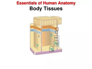
Human Tissues
Human Tissues. Anatomy and Physiology. Four types of tissues. Epithelial Connective Muscle Nerve. Simple squamous epithelium. "top" view. cells are large, but quite thin have a prominent, protruding nucleus (B). shape is the sunny-side-up fried egg.


Human Tissues
E N D
Presentation Transcript
Human Tissues Anatomy and Physiology
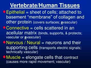
Four types of tissues Epithelial Connective Muscle Nerve
Simple squamous epithelium • "top" view. • cells are large, but quite thin • have a prominent, protruding nucleus (B). • shape is the sunny-side-up fried egg. • It has also been called "pavement epithelium, because it can look like like paving stones as seen from above
Simple cuboidal epithelium • Each kidney duct is lined by simple cuboidal epithelium • Edges generally aren't apparent, but whose nuclei are quite prominent • Each cell is almost a perfect cube - a square in cross section, as seen here
www.uoguelph.ca/zoology/devobio/ 210labs/epithelial1.html Simple columnar epithelium • From the small intestine • tall, vertical cells top of the tissue are columnar cells (one is shown - red arrow • cells at the bottom of the image are connective tissue, which will be discussed later • columnar cells are quite thick, they do not readily allow passive diffusion. • have numerous microvilli on their apical (lumenal) surface • the black arrow is indicating a goblet cell
Pseudostratified columnar epithelium single layer of cells of different lengths, giving the appearance of a multi layer structure
Stratified squamous epithelium • Stratified squamous epithelium is made up of multiple layers of flat cells. • This tissue makes up the outer layer of the skin, as well as the lining of the mouth and vagina.
Transitional epithelium • Note that the cells at the exposed surface (A) are large and rounded. They are also larger than the cells at the attached surface (B) • Locations: ureter, urinary bladderFunction: distention
Connective tissue • Functions mainly to bind and support other tissues. • They contain a few cells surrounded by a nonliving matrix. • This matrix can be fibrous, liquid or jellylike
Bone tissue • Bones are composed of mineralized connective tissue. • Cells called osteocytes deposit a matrix of collagen and calcium-phosphate. • Mammalian bone is constructed from repeated units called Haversian Units. • The process of making new bone is called ossification.
Compact bone tissue • In the center of the osteon is the central canal (A) which hold the blood vessels and a nerve. • These canals are surrounded by concentric rings of inorganic matrix,the lamellae (B). • Between the lamellae are bone cells, the osteocytes (C) located in lacunae. • Nutrients diffuse from cell to cell through the canaliculi (D).
Compact bone tissue A. Central canal B. Lamellae C. Osteocyte in lacunae D. Canaliculi
Diaphysis - shaft of bone • Epiphyses - ends of bone • Articular cartilage - covers epiphysis at a joint • Periosteum - membrane covers outer bone surface • Endosteum - lines medullary cavity • www.bmb.psu.edu/courses/ bisci004a/bone/bone.htm Parts of a bone
Functions of skeletal system 1. Support 2. Assist movement 3. Mineral "bank" 4. Blood cell production - hemopoesis (red marrow) 5. Energy storage - adipose tissue (yellow marrow)
Cartilage • Collagenous fibers embedded in a rubbery ground-substance called Chondrin, which is a protein-carbohydrate complex. • The chondron is secreted by chondrocytes. • Three types: hyaline, elastic, fibrocartilage
Hyaline cartilage • the bracket indicates the hyaline cartilage • Locations: "C" rings in the trachea, nose, articular ends of bones, fetal skeletonFunction: precursor to bone, support
Hyaline cartilage • This tissue is more highly magnified hyaline • The chondrocytes (A) are located in lacunae (C) • The matrix (B) contain collagen fibers that are so fine they are not visible in tissue preparations
Elastic cartilage Elastic cartilage is contained within the bracket The chondrocytes (A) are contained in lacunae (C). The matrix (B) contains abundant elastic fibers.These fibers give great flexibility to this tissue.
Elastic cartilage • The chondrocytes (A) are contained in lacunae (C). • The matrix (B) contains abundant elastic fibers. • These fibers give great flexibility to this tissue. • Locations: ear, auditory canal, epiglottisFunctions: flexible support
Fibrocartilage • Fibrocartilage is found in the area between the parallel lines • Locations: pubic symphysis, intervertebral discs • Functions: supports, withstands compression
Fibrocartilage • This cartilage type is recognized by chondrocytes (A) oriented in rows. • Highly magnified lacunae that hold the chondrocytes, are not visible. • The matrix (B) contains numerous fine collagen fibers. These fibers give the tissue durability.
Loose areolar connective tissue • Areolar tissue is a type of loose connective tissue. • Visible are the fibrous proteins giving this tissue its characteristic spaces. • Areolar refers to "space".
Areolar connective tissue • Nuclei of fibroblasts (A), collagen fibers (B) and elastic fibers (C). • Locations: beneath the skin and around blood vessels, muscles and nerves • Functions: binds one tissue to another (as skin connects to muscle), protection and nourishment to the organs and structures it binds, and stores "body fluid"
Fibrous Connective Tissue • Dense tissue owing to the large amounts of collagenous tissue. • Tendons and ligaments are the two types of structures containing this tissue.
Dense regular tissue • Dense regular connective tissue is found in tendons and ligaments. • The collagen fibers visible here give it strength when pulled. • fibroblasts (A) are more clearly observed between the parallel collagenous fibers (B).
Loose Connective Tissue • It binds epithelia to the underlying tissue and acts as a packing material to hold organs in place. • This type of tissue receives its name from the fibers it contains: collagenous, elastic, and reticular
Elastic Connective Tremendous number of elastic fibers (A). The fibroblasts are not visible. The light pink in this tissue is smooth muscle. Location: large arteries, bronchial tubes Function: Elastic fibers can stretch l 1/2 times their length and then recoil. These fibers will provide elasticity to tissues
Reticular connective • The reticular fibers (A) form a network or lattice in this spleen tissue. Do not confuse this tissue with the elastic connective tissue seen above which has fibers that are parallel. • Locations: spleen, lymph nodes, liver • Function: gives support to soft organs
Adipose Tissue • Loose connective tissue that stores fat. • These cells form when we are babies and fill and empty as we go through life. – • Anywhere there is an empty space in the body fat is stored as a source of energy and may provide insulation.
Adipose tissue • Observe that the nucleus (A) is pushed to the side of the cell giving the cell the appearance of a signet ring. Cells are filled with fat globules (B). • Functions:--The kidneys are correctly positioned and cushioned by adipose tissue.--The eye is cushioned in the orbit by adipose.
Blood tissue • Visible are the erythrocytes or red blood cells • A few cells seen are platelets
Blood cells Leukocytes- white blood cells are nucleated, larger than the red blood cells, which are non-nucleated
Muscles are • Composed of long contractile cells. The cells are composed of long microfilaments made of the proteins actin and myosin. • There are 3 types of muscle tissue: Striated or skeletal, cardiac, and visceral or smooth.
Muscles Voluntary Skeletal (striated) Involuntary Smooth Cardiac
Striated muscle Notice the striated appearance of the tissue and the elongated cells
Smooth muscle Notice the lumen in these images. The lumen is formed by epithelial tissue, but the adjacent layer is smooth muscle, which functions primarily to constrict or dilate the lumen.
Cardiac muscle Notice the striated appearance of the tissue, the intercalated discs, and the branching cells.
Nerve tissue • Senses stimuli and sends signals from one part of the body to another. The cells are called neurons. The neuron consists of a cyton (cell body), dendrites (many branches that carry impulses to the cyton), and an axon (long filament that carries impulses away from the cell).
Neuron 1. cell body (soma) is the factory of the neuron 3. dendrites receive signals from other nerve cells. 4. axon is the main conducting unit of the neuron
Synapse • Synapses are the junctions formed with other nerve cells where the presynaptic terminal of one cell comes into 'contact' with the postsynaptic membrane of another. It is at these junctions that neurons are excited, inhibited, or modulated. There are two types of synapse, electrical and chemical.
www.sunyniagara.cc.ny.us/ val/pscchigh.html www.gen.umn.edu/.../wa_histology/wa_epithelial_tissue.html References www.gen.umn.edu/faculty_staff/jensen/1135/webanatomy/ www.unomaha.edu/~swick/2740cartilagebone.html www.cnas.smsu.edu/labimages/ Biology/Bio122/week8.htm