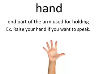
Hand
Hand. 19 Bones 19 Articulations 29 Muscles. Bones of the Hands. Arches of the Hand. Transverse carpal arch Transverse metacarpal arch Longitudinal arch. Mobility of 4 th and 5 th CMC Joints. Creases of the Hand. Distal digital crease Middle digital crease Proximal digital crease


Hand
E N D
Presentation Transcript
Hand 19 Bones 19 Articulations 29 Muscles
Arches of the Hand • Transverse carpal arch • Transverse metacarpal arch • Longitudinal arch
Creases of the Hand • Distal digital crease • Middle digital crease • Proximal digital crease • Distal palmar crease • Proximal palmar crease • Thenar crease • Distal wrist crease • Proximal wrist crease
Volar or Palmar Plates • Volar or Palmar Plates are dense thick discs of fibrocartilage which help to strengthen joint and prevent hyperextension • Note the fibrous digital sheath in top picture (annual pulley)
Flexion and Extension • Axis - Lateral • Plane - Sagittal • Abduction and Adduction • Axis - Anterior/Posterior • Plane – Frontal Motions at the MP Joints
Flexion and Extension • Axis - Lateral • Plane - Sagittal Motions at the PIP and DIP Joints
Muscles originating outside the hand • Flexor Digitorium Superficialis • Flexor Digitiorium Profundus • Flexor Pollicus Longus • Extensor Digitorum • Extensor Indicis Proprius • Extensor Digiti Minimi • Extensor Pollicus Longus • Extensor Pollicus Brevis • Abductor Pollicus Longus Extrinsics
Four Lumbricals • Three Palmar Interossei • Four Dorsal Interossei • Thenar muscles • Opponens Pollicus • Abductor Pollicus Brevis • Adductor Pollicus • Flexor Pollicus Brevis Intrinsics
Hypothenar muscles • Opponens Digiti Minimi • Abductor Digiti Minimi • Flexor Digiti Minimi Brevis • Palmaris Brevis Intrinsics
Flexor Digitorum SuperficialisTest for Tendon Integrity • Therapist holds all fingers except one being tested in extension. This isolates the Flexor Digitorum Superficialis. If client can flex at PIP joint then FDS tendon is intact.
Flexor Digitorum ProfundusTest for Tendon Integrity • Therapist extends all joints of client’s finger except the DIP. Therapist asks client to flex the DIP. If client can, FDP is intact
Annular Pulleys • Hold flexor tendons relatively close to joint (functional insertions) • Rupture results in bowstringing with less ROM and strength • Trigger finger
Over the proximal phalanx the extensor tendon (from extensor digitorum) divides into a central band and two lateral bands • The central band inserts at the base of the middle phalanx • The two lateral bands rejoin over the middle phalanx and insert at the base of the distal phalanx Extensor Assembly
MCP 70 degrees PIP/DIP extension Extensor MechanismClosed pack position
Relationship of AB & Adduction to Flexion and Extension at MP Joints • When MP joints are extended – the collateral ligaments are slack and allow for AB and Adduction of Fingers • When MP joints are flexed – the collateral ligaments are taut (tight) and prevent AB and ADduction
Position for Long Term Immobilization • Metacarpalphalangeal joints in 60 to 70 degrees of flexion • PIP and DIP joints extended
Thumb Flexion/Extension (Radial Adduction/Abduction) • Axis - Anterior/Posterior • Plane – Frontal • Thumb Palmar Adduction/Abduction • Axis – Lateral • Plane - Sagittal Thumb Movements at CMC Joint
Thumb Movements at CMC Joint • Flexion/Extension • (Radial AB/Adduction) • AB/Adduction • (Palmar AB/Adduction) • Opposition/Reposition
Functional Position of Hand • Wrist is in 20 to 30 degrees of extension and slight ulnar deviation • Fingers in 45 degrees of MCP, 15 degrees of PIP and DIP flexion • Thumb is in 45 degrees of abduction
Intrinsic Plus • Flexion of MP to 90 degrees and extension at PIP and DIP - or Roof Top Position • Interossei and lumbricals at their shortest • Common in patients with R.A.
Intrinsic Minus • Hyperextension of the MP joints and flexion of the PIP joints or “Clawhand” • Paralysis of interossei and lumbrical muscles
Intrinsic=(Lumbricals and interosseus =table top) • Extrinsic=ED, FDS, FDP) = Hook Intrinsic and extrinsicplus hand
Power grip • Spherical • Cylindrical • Precision grip • Power (key) pinch • Lateral pinch • Precision pinch • Hook grip Types of Prehension
Power grip • Spherical • Cylindrical • Precision grip • Power (key) pinch • Lateral pinch • Precision pinch • Hook grip Match
Common hand disorders
Intrinsic Tightness • Nerve injuries • Ulnar Nerve Injury • Median Nerve Injury • Carpal Tunnel Syndrome • Radial Nerve Injury • Tendon injuries • Mallet Finger • Swan Neck Deformity • Boutonniere Deformity • Zig Zag Deformities • DeQuervain’s Disease • Fascia • Dupuytren’s Contracture Problems of the Hand
MCP joint held in slight extension while examiner moves the PIP joint into flexion – if can’t be flexed, intrinsic or joint capsule tightness • Place MCP joint in a few degrees of flexion to relax intrinsics – if joint can now flex, then it was intrinsic tightness • If when MCP joint placed in flexion still can’t flex PIP – then it is a joint capsule tightness or contracture. Bunnell-Lister Test for Intrinsic Tightness
Bunnell-Lister Test for Intrinsic Tightness: Step 1 • MCP joint held in slight extension will therapist moves the PIP joint into flexion – if can’t be flexed, intrinsic or joint capsule tightness
Bunnell-Lister Test for Intrinsic Tightness: Step 2 • Place MCP joint in a few degrees of flexion to relax intrinsics – if joint can now flex, then it was intrinsic tightness
Bunnell-Lister Test for Intrinsic Tightness: Step 3 • If when MCP joint placed in flexion still can’t flex PIP – then it is a joint capsule tightness or contracture
1.) Coracoid process (may be injured during surgery) • 2.) Coracobrachialis muscle • 3.) Distal lateral arm as it goes through investing fascia • 4.) Lateral Forearm – Vulnerable to blunt trauma Musculotaneous nerve (C5, C6 – Continuation of the lateral cord)Points of entrapment
http://video.google.com/videosearch?sourceid=navclient&rlz=1T4ADBF_enUS296US296&q=tenodesis&um=1&ie=UTF-8&sa=N&hl=en&tab=wv#q=quadriplegia+c6&hl=en&emb=0http://video.google.com/videosearch?sourceid=navclient&rlz=1T4ADBF_enUS296US296&q=tenodesis&um=1&ie=UTF-8&sa=N&hl=en&tab=wv#q=quadriplegia+c6&hl=en&emb=0 Tenodesis- C6
Unable to oppose thumb • Unable to make a complete fist • Atrophy of thenar eminence • Weak wrist flexion • Weak pronation of the forearm Median Nerve Injury
1.) Ligament of struthers/supracondylar process (medial ridge) 2.) Bicipital aponeurosis 3.) Between 2 heads of pronator teres (Pronator syndrome) 4.) Sublimis Bridge (FDS borders) 5.) AIN (Anterior interosseous nerve branch)- may also be entrapped by pronator 6.) Carpal Tunnel- between flexor tendons and transverse carpal ligament 7.) Metacarpal tunnel – between metacarpal ligaments and MCP’s Median Nerve = C5-C6, Medial and Lateral cords
Flexor Carpi Radialis • Palmaris Longus • Flexor Digitorum Superficialis • Radial Half of Flexor Digitorum Profundus • Two Radial Lumbricals • Flexor Pollicus Longus • Superficial portion of Flexor Pollicus Brevis • Opponens Pollicus • Abductor Pollicus Brevis (may have ulnar innervation) Muscles Innervated by the Median Nerve
Carpal Tunnel Syndrome – Tinel’s Sign • Tinel’s Sign – When therapist taps over the carpal tunnel, the client will feel parasthesias or tingling distally
Phalen’s Test • Therapist flexes client’s wrists manually and holds together for one minute. Positive test elicits tingling in thumb, index finger, and middle and lateral half of the ring finger and is indicative of Carpal Tunnel Syndrome.
Low injury = Thumb, index, middle. Loss of 2 lateral lumbricals • Index and middle have noticeable claw, • Thumb is rotated and flexed and in same plane as fingers, looses opposition (ape) High injury = Only FCU and ulnar half of FDP are spared. Similar claw but not as pronounced because don’t have the force of the long flexors. (pope) Hand is virtually useless Median Nerve Injury (ape or pope)