Types of Ionizing Radiation
470 likes | 1.57k Vues
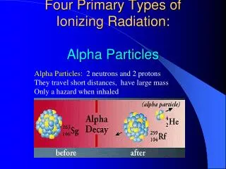
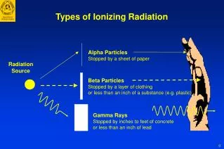
Types of Ionizing Radiation. Alpha Particles Stopped by a sheet of paper. Radiation Source. Beta Particles Stopped by a layer of clothing or less than an inch of a substance (e.g. plastic). Gamma Rays Stopped by inches to feet of concrete or less than an inch of lead. Radiation Units.

Types of Ionizing Radiation
E N D
Presentation Transcript
Types of Ionizing Radiation Alpha Particles Stopped by a sheet of paper Radiation Source Beta Particles Stopped by a layer of clothing or less than an inch of a substance (e.g. plastic) Gamma Rays Stopped by inches to feet of concrete or less than an inch of lead
Radiation Units Measure of Amount of radioactive material Ionization in air Absorbed energy per mass Absorbed dose weighted by type of radiation Quantity Activity Exposure Absorbed Dose Dose Equivalent Unit curie (Ci) roentgen (R) rad rem For most types of radiation 1 R 1 rad 1 rem
Radiation Doses and Dose Limits Flight from Los Angeles to London 5 mrem Annual public dose limit 100 mrem Annual natural background 300 mrem Fetal dose limit 500 mrem Barium enema 870 mrem Annual radiation worker dose limit 5,000 mrem Heart catheterization (skin dose) 26,000 mrem Life saving actions guidance (NCRP-116) 50,000 mrem Mild acute radiation syndrome 200,000 mrem LD50/60 for humans (bone marrow dose) 350,000 mrem Radiation therapy (localized & fractionated) 6,000,000 mrem
Radioactive Material • Radioactive material consists of atoms with unstable nuclei • The atoms spontaneously change (decay) to more stable forms and emit radiation • A person who is contaminated has radioactive material on their skin or inside their body (e.g., inhalation, ingestion or wound contamination)
Half-Life (HL) • Physical Half-Life Time (in minutes, hours, days or years) required for the activity of a radioactive material to decrease by one half due to radioactive decay • Biological Half-Life Time required for the body to eliminate half of the radioactive material (depends on the chemical form) • Effective Half-Life The net effect of the combination of the physical & biological half-lives in removing the radioactive material from the body • Half-lives range from fractions of seconds to millions of years • 1 HL = 50% 2 HL = 25% 3 HL = 12.5%
Examples of Radioactive Materials Physical RadionuclideHalf-LifeActivityUse Cesium-137* 30 yrs 1.5x10⁶ Ci Food Irradiator Cobalt-60 5 yrs 15,000 Ci Cancer Therapy Plutonium-23924,000 yrs 600 Ci Nuclear Weapon Iridium-192 74 days 100 Ci Industrial Radiography Hydrogen-3 12 yrs 12 Ci Exit Signs Strontium-90 29 yrs 0.1 Ci Eye Therapy Device Iodine-131 8 days 0.015 Ci Nuclear Medicine Therapy Technetium-99m 6 hrs 0.025 Ci Diagnostic Imaging Americium-241 432 yrs 0.000005 Ci Smoke Detectors Radon-222 4 days 1 pCi/l Environmental Level *Potential use in radiological dispersion device
Types of Radiation Hazards Internal Contamination • External Exposure - whole-body or partial-body (no radiation hazard to EMS staff) • Contaminated- • external radioactive material: on the skin • internal radioactive material: inhaled, swallowed, absorbed through skin or wounds External Contamination External Exposure
Causes of Radiation Exposure/Contamination • Accidents • Nuclear reactor • Medical radiation therapy • Industrial irradiator • Lost/stolen medical or industrial radioactive sources • Transportation • Terrorist Event • Radiological dispersal device (dirty bomb) • Attack on or sabotage of a nuclear facility • Low yield nuclear weapon
Scope of Event Event Number of Deaths Most Deaths Due to Radiation None/Few Radiation Accident Few/Moderate Radioactive Blast Trauma (Depends on Dispersal size of explosion & Device proximity of persons) Blast Trauma Low Yield Large Thermal Burns (e.g. tens of thousands in NuclearWeapon an urban area even from Radiation Exposure 0.1 kT weapon) Fallout (Depends on Distance)
Radiation ProtectionReducing Radiation Exposure • Time • Minimize time spent near radiation sources To Limit Caregiver Dose to 5 rem Distance Rate Stay time 1 ft 12.5 R/hr 24 min 2 ft 3.1 R/hr 1.6 hr 5 ft 0.5 R/hr 10 hr 8 ft 0.2 R/hr 25 hr • Distance • Maintain maximal practical distance from radiation source • Shielding • Place radioactive sources in a lead container
Detecting and Measuring Radiation • Instruments • Locate contamination - GM Survey Meter (Geiger counter) • Measure exposure rate - Ion Chamber • Personal Dosimeters - measure doses to staff • Radiation Badge - Film/TLD • Self reading dosimeter (analog & digital)
Patient Management - Decontamination • Carefully remove and bag patient’s clothing and personal belongings (typically removes 95% of contamination) • Survey patient and, if practical, collect samples • Handle foreign objects with care until proven non-radioactive with survey meter • Decontamination priorities: • Decontaminate wounds first, then intact skin • Start with highest levels of contamination • Change outer gloves frequently to minimize spread of contamination
Patient Management - Decontamination (Cont.) • Protect non-contaminated wounds with waterproof dressings • Contaminated wounds: • Irrigate and gently scrub with surgical sponge • Extend wound debridement for removal of contamination only in extreme cases and upon expert advice • Avoid overly aggressive decontamination • Change dressings frequently • Decontaminate intact skin and hair by washing with soap & water • Remove stubborn contamination on hair by cutting with scissors or electric clippers • Promote sweating • Use survey meter to monitor progress of decontamination
Patient Management - Decontamination (Cont.) • Cease decontamination of skin and wounds • When the area is less than twice background, or • When there is no significant reduction between decon efforts, and • Before intact skin becomes abraded. • Contaminated thermal burns • Gently rinse. Washing may increase severity of injury. • Additional contamination will be removed when dressings are changed. • Do not delay surgery or other necessary medical procedures or exams…residual contamination can be controlled.
Treatment of Internal Contamination • Radionuclide-specific • Most effective when administered early • May need to act on preliminary information • NCRP Report No. 65, Management of Persons Accidentally Contaminated with Radionuclides RadionuclideTreatment Route Cesium-137 Prussian blue Oral Iodine-125/131 Potassium iodide Oral Strontium-90 Aluminum phosphate Oral Americium-241/ Ca- and Zn-DTPA IV infusion, Plutonium-239/ nebulizer Cobalt-60
Facility Recovery • Remove waste from the Emergency Department and triage area • Survey facility for contamination • Decontaminate as necessary • Normal cleaning routines (mop, strip waxed floors) typically very effective • Periodically reassess contamination levels • Replace furniture, floor tiles, etc. that cannot be adequately decontaminated • Decontamination Goal: Less than twice normal background…higher levels may be acceptable
Radiation Sickness Acute Radiation Syndrome • Occurs only in patients who have received very high radiation doses (greater than approximately 100 rem) to most of the body • Dose ~ 15 rem • no symptoms, possible chromosomal aberrations • Dose ~ 50 rem • no symptoms, minor decreases in white cells and platelets
Acute Radiation Syndrome (Cont.)For Doses > 100 rem • Prodromal stage • nausea, vomiting, diarrhea and fatigue • higher doses produce more rapid onset and greater severity • Latent period (Interval) • patient appears to recover • decreases with increasing dose • Manifest Illness Stage • Hematopoietic • Gastrointestinal • CNS Time of Onset Severity of Effect
Acute Radiation Syndrome (Cont.)Hematopoietic Component - latent period from weeks to days • Dose ~ 100 rem • ~10% exhibit nausea and vomiting within 48 hr • mildly depressed blood counts • Dose ~ 350 rem • ~90% exhibit nausea/vomiting within 12 hr, 10% exhibit diarrhea within 8 hr • severe bone marrow depression • ~50% mortality without supportive care • Dose ~ 500 rem • ~50% mortality with supportive care • Dose ~ 1000 rem • 90-100% mortality despite supportive care
Acute Radiation Syndrome (Cont.)Gastrointestinal and CNS Components • Dose > 1000 rem - damage to GI system • severe nausea, vomiting and diarrhea (within minutes) • short latent period (days to hours) • usually fatal in weeks to days • Dose > 3,000 rem - damage to CNS • vomiting, diarrhea, confusion, severe hypotension within minutes • collapse of cardiovascular and CNS • fatal within 24 to 72 hours
Treatment of Large External Exposures • Estimating the severity of radiation injury is difficult. • Signs and symptoms (N,V,D,F): Rapid onset and greater severity indicate higher doses. Can be psychosomatic. • CBC with absolute lymphocyte count • Chromosomal analysis of lymphocytes (requires special lab) • Treat symptomatically. Prevention and management of infection is the primary objective. • Hematopoietic growth factors, e.g., GM-CSF, G-CSF (24-48 hr) • Irradiated blood products • Antibiotics/reverse isolation • Electrolytes • Seek the guidance of experts. • Radiation Emergency Assistance Center/ Training Site (REAC/TS) • Medical Radiobiology Advisory Team (MRAT)
Localized Radiation Effects - Organ System Threshold Effects • Skin - No visible injuries < 100 rem • Main erythema, epilation >500 rem • Moist desquamation >1,800 rem • Ulceration/Necrosis >2,400 rem • Cataracts • Acute exposure >200 rem • Chronic exposure >600 rem • Permanent Sterility • Female >250 rem • Male >350 rem
Emergency Surgery Hematopoietic Recovery No Surgery Surgery Permitted 24 - 48 Hours After adequate hematopoietic recovery ~3 Months Special Considerations • High radiation dose and trauma interact synergistically to increase mortality • Close wounds on patients with doses > 100 rem • Wound, burn care and surgery should be done in the first 48 hours or delayed for 2 to 3 months (> 100 rem)
Chronic Health Effects from Radiation • Radiation is a weak carcinogen at low doses • No unique effects (type, latency, pathology) • Natural incidence of cancer ~ 40%; mortality ~ 25% • Risk of fatal cancer is estimated as ~ 5% per 100 rem • A dose of 5 rem increases the risk of fatal cancer by ~ 0.25% • A dose of 25 rem increases the risk of fatal cancer by ~ 1.25%
What are the Risks to Future Children?Hereditary Effects • Magnitude of hereditary risk per rem is ~10% that of fatal cancer risk • Risk to caregivers who would likely receive low doses is very small - 5 rem increases the risk of severe hereditary effects by ~ 0.02% • Risk of severe hereditary effects to a patient population receiving high doses is estimated as ~ 0.4% per 100 rem
Fetal IrradiationNo significant risk of adverse developmental effects below 10 rem Weeks After Fertilization Period of Development Effects <2 2-7 7-40 All Pre-implantation Organogenesis Fetal • Little chance of malformation • Most probable effect, if any, is death of embryo • Reduced lethal effects • Teratogenic effects • Growth retardation • Impaired mental ability • Growth retardation with higher doses • Increased childhood cancer risk (~ 0.6% per 10 rem)