Download
1 / 226
2.26k likes | 2.42k Vues
Explore the anatomy, histology, physiology, and functions of the G.I. tract and its accessory organs in the digestion process. Learn about the essential roles of organs like the mouth, stomach, pancreas, liver, gallbladder, and small intestine in nutrient breakdown and absorption. Understand the importance of gastrointestinal hormones, movements of the G.I. tract, and absorption processes in maintaining overall health and energy production from food. Dive into the complexities of the human digestive system and how it processes food for energy and tissue repair.

E N D
1) INTRODUCTION OF G.I. TRACT. • Function. • Anatomical consideration. • Histological structure. • Blood supply. • Nerve supply. 2) Different Organs Involved in Process of Digestion. • Mouth. • introduction. • General Physiological, action • Salivary Glands. • Apllied Physiology.
b) Tongue c) Teeth d) Pharynx e) Esophagus • Introduction • Histology • Physiology • Applied f) Stomach • Introducton • Functional Anatomy • Histology • Gland of stomach
v)Function of Stomach vi) Applied physiology g) Pancreas • Anatomy • Histology iii) Pancreatic juice iv) Applied physiology h) Liver and Gallbladder • Introduction • Anatomy of Liver • Billiary system • Blood supply • Composition of Bile
vi) Properties vii) Storage of Bile viii) Function of bile ix) Function of Liver Gallbladder • Introduction • Function of Gallbladder. • Applied physiology of Gallbladder and Liver. • Small Intestine • Introduction • Anatomy • Histology • Properties and composition of succusentericus
v) Function of succus entericus. vi) Function of small Intestine. J) Gastrointestinal Hormones. K) Movements of G.I.T L) Absorptions
INTRODUCTION The food we eat contains a variety of nutrients, which are used for building new body tissues and repairing damaged tissue. Food is also vital for life because it is our only source of chemical energy. As consumed however, most food cannot be used either to build tissues or to power the energy requiring chemical reaction of body calls. Rather, food must be broken down into molecule that are small enough to enter body cells, a process known as Digestion. The passage of these smaller molecules through the plasma membranes of cells lining the stomach and Intestines and then into the blood and lymph is termed absorption. The organs that collectively perform there function are collectively make a system known as Digestive System.
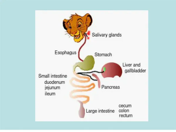
Two groups of organs compose the digestive system :- 1 The gastrointestinal tract 2 accessory digestive Organs The gastrointertinal tract or alimentary canal (alimentary-nourishment), is a continous tube that extends from the mouth to anus organs if the gastrointestinal tract include the • Mouth • Must if the pharynx • esophagus
Stomach • Small Intestine • And Large Intestine The length of GI tract taken from a cadaver is about 9m (30 Ft). In a living person, it is much shorter because the muscles along the walls of G.I. tract organs are in a state of tonus (sustained contraction). The accessony digestive organs are as follows:- • Teeth • Tongue • Salivary glands • Liver • Gall bladder • And pancreas.
Teeth aid in physical breakdown of food and the tongue assist in chewing and swallowing. The other accessory digestive organs hour however never come into direct contact with food. They produce on store secretions that flow into the GI tract through ducts.: the secretion aid in the chemical breakdown of food . The GI tract contains and processes food from the time it is eaten until it is digested and absorbed on eliminated. In some parts of the GI tract muscular contraction in the wall physically break down the food by churning it. The contractions also help to dissolve food by mixing them with fluids secreted into the tract. Enzymes secreted by accessory structures and cells that line the tract break down the food chemically. Wave line contractions of the smooth muscle in the wall of the smooth muscle in the wall of the GI tract propel the food along the tract from the esophagus to the anus.
Overall, the digestive system Perform six basic process:- • Ingestion:- This process involves taking foods and liquids into the mouth i.e eating. • Secretion:- Each day, cells within the walls of the GI tract and accessory digestive organs secret a total of about 7 liters of water, acid, buffers and enzymes into the lumen of the tract. 3) Mixing and Propulsion:- Altering contraction and wall relaxation of smooth muscle in the walls of the GI tract mix food and secreations and propel them towards the anus. This capability of the GI tract to mix and move material along its length is termed motility.
4) Digestion:- Mechanical and chemical processes break down ingested food into small molecules. In mechanical digestion the teeth cut and grind food before it is swallowed and then smooth muscles of the stomach and small intestine churn the food. As a result, food molecules become dissolved and thoroughly mixed with digestive enzymes. In chemical digestion the large carbohydrate lipid, protein and nucleic acid molecules in food are split into smaller molecules by hydrolysis. Digestive enzymes produced by the salivary glands , tongue, stomach, pancrease and small intestine catalyze these catabolic reaction
. A few substances in food can be absorbed without chemical digestion. These include amino acids, cholestral, glucose, vitamins, minerals and water. 5) Absorption:- The entrance of ingested and secreted fluids, ions and the small molecules that are products of digestion into the epithelial cells lining the lumen of the GI tract is called absorption. The absorbed substances pass into blood or lymph and circulate to cells throughtout the body. 6) Defection:- Wastes, indigestible substances bacteria cells sloughed from the lining of the GI tract and digested materials that were not absorbed leave the body through the anus in a process called defection. The eliminated material is termed feces.
Anatomical Consideration The human digestive canal is a large muscular tube consist of the following parts from above downwards the mouth (guarded by lips and teeth), tongue, pharynx, esophagus , stomach, small intestine, large intestine, rectum and anal canal. The ducts of the salivary glands open into the mouth. The proximal end of the stomach (i.e. its junction with oesophagus) is guarded by the cardiac sphineter. The distal end of the stomach is guarded by the pyloric sphineter. The small intestine begins after the pyloric sphincter and consists successively of the following subdivisions. Duodenum, jejunum and ileum. The deodenum receives food from the stomach. The bile duct and pancreatic duet jointly open in it through the ampula of vater.
The small intestine is very long and is roughly about 7.6 meters (25 feet). The great length of small intestine provide enough time and surface area so that digestion and absorption of food stuff may be complete. The small intestine opens into the next part the large intestine. The opening between them is guarded by iliocolic sphincter. In the LI water is absorbed and the faeces become formed. The LI opens into the last part rectum and anal canal. Histological Structure The wall of the aliminatery canal from the oesophagus to the anal canal consist of four encircling ayers or tunics from outside to inwards. 1) Tunica adventitia or serosa:- Fibrous outer coat carries large blood vessels and nerves. When the organ is covered by the peritoneum, this layer is called serosa, otherwise tunica adventitia.
2) Tunica muscularis:- is consist of a double layer of smooth muscle of which the inner sheet lies circularly and the outer sheet run longitudinally. Nerve and vascular plexuses lie between the layers. This muscular layer controls the diameter of the intestine and propels its contents towards the anus. 3) Tunica submucosa:- is a loose arealer connective tissue layer which contains large blood and lymphatic vessels nerves and glands in certain portions. 4) Tunica mucosa:- mucousa membrane consist of i) stratified squamous or single columnar epithilial layer. ii) Laminopropria which is a delicate areolar connective tissue layer. iii) Muscularis mucosa is a thin layer of smooth muscle and forms the boundary between mucous membrane and sub mucosa.
Blood supply:- The gastr-intestinal circulation is the largest systemic regional vascular bed. In resting conditions only one-third of the cardiac output flows through the abdominal organs. Anatomically, the gastrointestinal circulation includes includes all the blood vessels supllying the salivary glands, mouth, phrynx, oesophagus, stomach, etc. However, the term gastro-intestinal circulation usually refers to vessels lying in the abdomen- often called the splanchinic circulation.
The liver receives one third of its of its blood through the hepatic artery and two-thirds through the portal vein. • (Appriximately 80% of the blood flow to the alimentary canal goes to the mucosa.
NERVE SUPPLY The nerve supply consist of:- • An intrinsic part:- which is represented by nerve and cells fibres originating and located in the Intestinal wall itself. • An intrinsic portion:- which is represented by vogal fibres of the symphatic. The intrinsic nueral structures supply the smooth muscularature of the alimenatary canal except the striated muscle fibres of the mouth, the upper part of the oesophagus and sphincter the intrinsic mechanism consist of a series of plexuses which are composed of small groups of nerve cells and bundle of nerve fibres. Two intramural plexus are located in a narrow space.
Between longitudinal and circular muscle layer s of the intestine wall myenteric plexus of Auerbach and. Between circular muscle layer and subcusa (submucosa plexus of meissner). This intrinsic networks of nerves within the walls of the digestive tube is responsible for the spontaneous movements of the intestinal tract even after the extrinsic network has been cut. The extrinsic nerve supply acts to tegulater the intrinsic neuromuscular mechanism that determines the movements of the digestive tube. However, the intrinsic neural mechanism is modulated by extrinsic nerve supplied by both of the autonomic nervous system. The sympathatic fibres which are branches of the splanchnic nerves. The sympathatic also appear to supply blood vessels within the intestinal wall. The preganglionic parasympathatic fibres are derived from:-
The cells in the medulla oblongata and The sacral segments of the spinal cord. These fibres originating from the medulla oblangata are mainly parts of vagal ones and supply muscles of trhe stomach, small intestine and upper half of large intestine. The pregangliolic fibres from the sacral spinal cord reach the pelvic ganglion by way of the pelvic nerves and postganglionic fibres supply the lower half of the LI end the rectum. The internal anal sphincter is innervated by efferent and somatic nerve fibres. The Sympathatic fibres are excitatory for ileocaecal and internal anal sphincters and smooth muscle fibres of the muscularies mucosa throughout the whole GI canal and help to increase the number of fields in the tract are inhibitory for the remaining musculative.
The parasympathetic system is excitatory for all the musculature except the sphinters where it is usually inhibitory. The two nervous system are antagonistic but so far as chyne is concerned. Function of the Digestive System:- The human digestive system serves the following function:- • Ingestion of the food • Digestion of food. • Secretion of various digestive juices. • Absorption of water, salts, vit, and end products of food digestion. • Excretion heavy metals, toxins certain alkaloids etc. • Movement certain types of movements are present in the whole of the GIT the functions of there movements are to facilitate admixture of food with digestive juices to propel food onwards to help blood and lymphatic circulation through the intestinal wall. Dafaecation also due to the movement of large intestine.
Mouth The mouth also referred to as the oral or buccal cavity is formed by the cheeks hard and soft palates and tongue forming the lat walls of the oral cavity are the cheeks. Then lips or labia are fleshy folds surrounding the opening of the mouth. The inner surface of each lip is attached to its corresponding gums by mid line fold of mucous membrane called labium franulum. During chewing contraction of the Buccinator muscle in the cheeks and orbiculariesoris muscles in the lips help keep food between the upper and lower teeth & also help in the speech. The vestibule of the oral cavity is a space bounded by the cheeks & lips & internally by the gums & teeth. The oral cavity proper is a space that extension from the gums and teeth to the fauces.
The opening between the oral cavity & pharynx. The hard palate the ant portion of the roof of the mouth is formed by the maxillae & palatine bones, is covered by mucous membrane. & forms a bony partition between the oral cavity & pharynx. The hard plate the ant portion of the roof of the mouth is formed by the maxillae & palatine bones is covered by mucous membrane, & forms partition between the oral and nasal cavities. The soft palate which forms the post portion of the roof of the mouth, is an arched muscular portion between the oropharynx and nasopharynx. Hanging from the free border of the soft palate is a conical muscular process called the uvula. lateral to base of uvula are two muscular folds than run down the lateral sides of the soft palate.
Antiriorly palatoglossial arch extends to the side of the base of the tongue. Postiorly palatopharageal arch extends to the side of the pharynx. The palatine tonsils are situated between the arches & the lingual are situated at the base of tongue. At the posterior border of soft palate the mouth open into the oropharynx through the fauces. General physiologically action of the mouth. The opening in the mouth through which food enters the body. Even before a person takes on initial bite of food the sensation provided by sight smell & even imagination of appetizing food prompts the salivary glands under the tongue to secret saliva. This prepares the body for the meal to come.
As the teeth grind the food into smaller bits saliva moistens it for easier swallowing. A digestive enzyme in the saliva c/a amylose breaks down carbohydrate (sugar & starch) muscle movement in the tongue & mouth provide the swallowing necessary to push the food into the throat (pharynx). This is a passage for food and air that s abut 5 inches long (12.7cm). A flexible flap of tissue called the epiglottis reflexively closes over the wind pipe during swallowing to prevent choking & to prevent food from entering the tongue. Salivary Glands:- A salivary is any cell or organ that releases a secretion called saliva to the oral cavity. The main function of saliva is to keep the mucous membrane of the mouth & pharynx moist & cleans the mouth & teeth.
When food enters the mouth secretion of saliva & it lubricates and begins chemical breakdown of food. The mucous membrne of mouth & and tongue contains many salivery glands that open directly or indirectly via short ducts to the oral cavity. This glands includes lapial, buccal & palatal glands in the lips, cheeks & palates respectively & lingular glands in the tongue all of which make a small contribution to saliva. however most saliva is secreted by the major salivery glands which lie beyond the oral mucosa. There are 3 pairs of major salivery glands. They are as under:- • parotid gland • Submedibular • Sublingual
Situation:- The parotidg. are located inf & ant to the ears between the skin &messater muscle. Each gland about 20-30gm in adults secretion from this glands are emptied into the oral cavity by stenson’s duct. This is opens inside cheek. Submedibular gland:- are found beneath the base of the tongue in part of the floor of the mouth. Each gland weight about 8-10 gm. Saliva from this gland empitit into the oral cavity by Wharton’s duct which is about 90 mm long. The sublingual gland are sup to submendi glands. This gland are smallest situated in mucous at the floor of the mouth. Each gland weight bout 2-3 gm. Saliva from this gland is poured into 5-15 small ducts called Ductus of ravinus.
This duct open on small papilae beneath the tongue among this duct one is longer called Barthalin’s duct which drains the ant part of the gland. There is some adjustments:- Composition:- Mixed saliva contain 99.57, water & 5% organic or inorganic substances. A part from these, gases are also found in sliva. Organic sub:- 1) Salivary proteins-Mucin & Albumin. 2) Salivary enzymes – Amylose, Maltose, Lysozyme, phosphatase. 3) Blood group components- Antigens 4) Free amino acids 5) Non protien nitrogens substances like urea, uric acid, creatinine, xanthine & hypoxanthine. Glucose is normally absent in saliva however in diabetic patients, glocose is found in sliva.
Inorganic sub:- 1) Sodium (Na) 2) ca 3) k 4) Bicarbonates 5) Bromide 6) chloride 7) flouride Gases present in saliva:-1) O2 2) Co2 3) N2 Functions of saliva:- Saliva perform following function:- • Prepration of food for swallowing:- As soon as food enters the mouth, saliva moistens & dissolves it. The mucous membrane of mouth is also moistened by saliva & thus facilitates chewing. By the movement of the tongue, the moistened & masticated food is rolled into the bolus. The mucin of saliva moistens & lubricates the a bolus & facilitates the swallowing.
2) Appreciation of taste:- Taste is a chemical sensation saliva by its solvent action dissolves the solid substance, so that these dissolved substances can stimulate the taste buds. 3) Digestive function:- Salivary amylase on cooked or bolied starch & convert it into maltase. Though starch digestion start in the mouth major part of it occurs in the stomach because the food stays only for a short time in the mouth. The optimum PH necessary for the activation of salivary amylase is 6 . The salivary amylase can’t act on cellulose. The enzyme maltase is present only traces in human saliva it convert maltose into glucose.
4) Cleansing and protective function:- Due to the constant secretion of saliva the mouth and teeth are rinsed and kept free from food debris and foreign particles. In this way saliva prevent growth of bacteria by removing materials which may serve as culture media for the growth of bacteria lysozyme of saliva kills so many bacteria like staphylococcus, streptococcus. 5) Role in speech:- By moistening and lubricating the soft parts of mouth and lips, saliva helps in speech. If the mouth is dry, articulation and pronounciation become difficult. 6) Excretory function:- Many substances both organic and inorganic are excreted in saliva.It excrete substances like mercury, potassium, iodide, lead. In some pathological condition , saliva excretes some substances like urea in nephritis and calcium hyperparathyroidism.
7) Regulation of body temperature.:- In dogs and cattle excessive dripping of saliva during panting helps in loss of heat. But in human being, saliva does not take part in temperature regulation. 8) Regulation of water balance :- When the body water content is reduced it decreases the salivary secretion also. This cause dryness of the mouth and induces thirst when the water is taken it quenches the thirst and restores the body water content. Secretion of saliva:- The daily secretion of saliva normally ranges between 800 to 1500 ml. saliva contains two major types of protein secretion:- 1) A serous secretion that contains ptyaline (an α amylase) which is an enzyme for digesting starches.
2) Mucous secretion:- that contains mucin for lubricating and for surface protective purposes. The parotid glands secrete entirely the serious type of secretion and the sub mandibular and sublingul glands secrete both the serous type and mucus. The buccal glands secrete only mucus. Saliva has a PH between 6.0 and 7.0 a favourable range for the digestive action of ptyalin. Regulation of salivary secretion:- Saliva is continuously secreted. During mastification of the food, the secretion is increased. The secretion of saliva is regulated only by nervous mechanism. Nerve supply to Salivary glands:- Salivary glands are under control of autonomic nervous system and receive efferent nerve fibres from both parasymathatic and sympathatic divisions of autonomic nervous system.
Reflex mechanism of salivary secretion:- The salivary secretion is a reflexes phenomenon salivary reflexes are of two types, namely unconditioned reflex and conditioned. Unconditioned Reflex:- When any substance is placed in the mouth the salivary secretion occurs. This is called the unconditional reflex and it is due to the stimulation of nerve endings in the mucus membrane of the oral cavity. This reflex is inborn and occurs immediately after birth. This quality of the substance is immaterial. Condition Reflex:- Salivary secretion also occurs by the sight, smell or thought of food. This reflex is called conditioned reflex.
This is due to the impulses arising from eyes. Nose etc. This is not inborn reflex but it is an acquired reflex. This need previous experience.
Applied Physiology Hyposalivation:- The reducation in the secretion of saliva is called the hyposalivation. It is of two types namely the temperoryhyposalivation and the permanent hyposalivation. • Temperoryhyposalivation occurs in the following condition:- • i) Emotional condition like fear, ii) Fever and iii) Dehydration b) Permanent hyposalivation occurs:- i) obstruction of salivary duct. • Aptyalism or xerostomia- congenital absence or hypoplasia of salivary glands and • Bell’s Palsy – paralysis of facial nerve
Hypersalivation or Sialorrhea:- The increase in the secretion of saliva is called hypersalivation or sialorrhea. This occurs in the following condition. • Pregnancy- due to unknown cause. • Decay of tooth or neoplasm of mouth or tongue due to continuous irritation of nerve ending in the mouth. • Disease of esophagus, stomach and intestine. • Some psychological condition • Nausea and vomiting. Chorda tympani syndrome:- During the regeneration of nerve fibres following trauma or surgical division, some of the nerve fibres of salivary gland which pass through chorda tympani branch of facial nerve may be misdirected and join with the nerve fibers supplying sweat glands.
So, when the food is taken in the mouth, salivary secretion is associated with sweat secretion. This is called the chorda tympani syndrome. Paralytic secretion of saliva:- When the parasympathatic nerve to salivary gland is cut, salivary secretion increases for 3 weekes and later diminishes and then stops at about 6th week. The increased secretion of saliva after cutting the parasympathatic nerve fibers is called paralytic secretion. This is because of release of more amount of andrenaline from the suprarenal glands after the denervation. The acinar cells of the salivary glands are hypersensitive to adrenaline. The paralytic secretion does not occur after cutting the sympathatic nerves fibers to salivary glands.
Mumps:- Although any of the salivary glands may be the target a nasophryngeal infections the mumps virus typically attacks the parotid glands. Mumps is a inflammation & enlagement of parotid gland accompanied by moderate fever, and extreme pain in the throat, especially when swallowing sour foods or acidic juices. Tongue:- The tongue is an accessory digstive organ composed of skeletal muscle covered with mucous membrane together with its associated muscle, it forms the floor of the oral cavity. The tongue is divided into symmatrical lateral halves by a median septum that extends its entire length and it is attached inferiorly to the hyoid bone, styloid process of temporal bone and mandible. Each half of the tongue consist of an identical complement of extrinsic and intrinsic muscle.
The extrinsic muscle of the tongue which originate outside the tongue & insert into the connective tissue in the tongue. The intrinsic muscle move the tongue from side to side and in and out to maneuver food for chewing and force the food to the back of the mouth for swallowing. They also form the floor of the mouth and hold the tongue in position. The intrinsic muscle alter the shape and size of the tongue for speech and swallowing. The lingual frenulum a fold of mucous membrane in the midline of the under surface of the tongue attached to the floor of the mouth and aids in limiting the movement of the tongue posteriorly. If a person’s lingual frenulum is abnormally short or rigid a condition called ankyloglossia then eating and speaking are impaired such that the person is said to be tongue tied.
The dorsum and lateral surface of the tongue are covered with papilliae projection of lamina propria covered kertinized epithilium. Many papillae contain taste buds, the receptors for gustation (taste) Fungiform papillae are mushroom like elevation distributed among the filiform papillae that are more numerous near the tip of the tongue. They appear as red dots on the surface of the tongue and most of them contain taste buds. Vallate papillae are arranged in an inverted v shaped on the posterior surface of the tongue all of them contain taste buds. Foliate papillae are located in small trenches on the lateral margins of the tongue but most of their taste buds degenerate in early childhood.
Filiform papillae are pointed thread like projections distributed in the parallel rows over the anterior two third of the tongue. Although filiform papillae lack taste buds they contain receptors for touch and increase friction between the tongue and food making it easier for the tongue to move food in the oral cavity. Lingual glands in the lamina propria secrete both mucus and a watery sereous fluid that contains the enzymes lingual lipase which acts on triglycerides. Teeth:- The teeth are accessory digestive organs located in sockets of the alveolar processes of the mandible and maxillae. A typical tooth has three major regions. The crown is the visible portion above the level of the gums.
PHARYNX It is musculo membraneous tube whose constricted end in the oesophagus. Its division from above downward are nasal, oral and laryngeal. The mucous membrane lines the pharynx. Its walls are provided with sensory receptors. These receptors are sensitive to mechanical stimulation and are essential in the mechanism of swallowing. When liquid are food stimualtes there touch receptors, a complex refelex of swallowing is initiated, but when there sensory areas are anaes thetised, swallowing becomes difficult.
Other than function of transmission of air from nose or mouth to the larynx and production of voice pharynx serves as a channel to transport food from mouth to oesophagus. During deglutition or swallowing, closure of mouth and nasopharynx effectively snuts of the pharynx from outside atmosphere. Dilation of closed pharynx by contraction of pharyngeal muscle result in development of a slight negative pressure when this effect is combined with thrust, food is pushed downward and onward in the oesophagus.