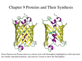
Understanding Protein Structure, Functions, and the Role of Amino Acids in Biological Catalysis
This chapter delves into the fascinating world of proteins and amino acids, highlighting their significance in biological systems. It covers the basic structure of amino acids, including their unique side chains, and explains the classification of amino acids based on polarity and acidity. Further, the chapter explores the hierarchical structure of proteins, from primary to tertiary, and emphasizes the importance of protein folding for biological function. Additionally, examples of globular and fibrous proteins, as well as disorders related to collagen malfunction, are discussed.

Understanding Protein Structure, Functions, and the Role of Amino Acids in Biological Catalysis
E N D
Presentation Transcript
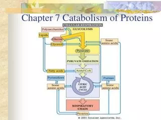
Chapter 9Proteins Amino Acids Protein Structure Protein Functions Enzymes—Life’s Catalysts
Amino Acids a carbon amino group carboxyl group side chain H O | || H2N – C – C – OH | R There are 20 different (genetically encoded) amino acids. These differ only in the nature of the side chain, R.
Amino Acids pK = 9.7 H O | || H2N – C – C – OH | R H O | || ↔ H2N – C – C – O- | R + H+ H O | || H3N+ – C – C – OH ↔ | R pK = 2.3 + H+ The amino group is a base and can accept a proton. The carboxylic acid group is an acid and can donate a proton Given that ‘R’ for the amino acid alanine is –CH3, Draw the structure of alanine. Given that the pK’s are 2.3 for the -COOH group and 9.7 for the –NH2 group, describe it’s ionic form at various pH values. What is it’s ionic form in blood at pH 7.4?
Amino Acid at ~ pH 7 H O | || H3N+ – C – C – O- | R Except for Glycine ( R = H) all amino acids have a chiral C. However only the ‘L’ form exists in nature. Natural Carbohydrate molecules (e.g. sugars) are …. a) always D b) always L c) either d) not chiral
Amino acids are typically classified based on the structure of the side chains as …… Nonpolar C and H atoms only (also Methionine – a thioether) Polar O and N in forms that cannot form ions (ionize) alcohols, amides acid form conjugate base Acidic COOH – carboxyl group - COOH ↔ -COO- + H+ base form conjugate acid Basic amino group (-NH2) H+ + NH2 ↔ NH3+ could be 2ndary amine but not amide
Nonpolar amino acids Could be considered polar or nonpolar
Combination of amino acids by a condensation reaction Dipeptide Tripeptides − oligo peptides − polypeptides A biologically active polypeptide containing 50 or more amino acids is a protein. Proteins may also contain more than one polypeptide chain (subunits). They must fold up into a specific, functional structure.
Aspartame, an artificial sweetener, is a modified dipeptide (at pH 7.4) H O | || H3N+ – C – C – | CH2 | COO- H O | || - N – C – C – O – CH3 H | CH2 |
Protein Structure Primary Structure The primary (1) structure of a protein is the order in which the amino acids are joined together to form the protein backbone. The side chains of the amino acids are substituents dangling from this backbone. The peptide bond is the covalent bond that forms this level of protein structure. Note that the backbone repeats ….. The amino and carboxy ends are typically charged at pH 7.4 carboxy end amino end
C = O ••• H – N Hydrogen bonding Protein Structure: Secondary Structure Backbone hydrogen bonding b-sheet extended structure zig-zag due to 109º or 120º a-helix Coiled structure Like spring
Protein Structure: Tertiary Structure Side chain interactions External side chains (polar/acidic/basic) can interact with water rather than another amino acid disulfide bonds covalent – 2 Cys only H-bonds 2 polar side chains Salt bridge 1 acidic + 1 basic side chain both need to be ionized (like ionic bond) Hydrophobic Nonpolar side chains Interactions inside of protein away from water (solvent) Protein Folding
Proteins are a cell’s tool kit. They each have a job to do (function) …… and a shape that is specific to the job they must perform. Protein Folding
Proteins are a cell’s tool kit. They each have a job to do (function) …… and a shape that is specific to the job they must perform.
Globular vs. Fibrous Proteins • Globular proteins fold into a compact, spherical shape with polar amino acid side chains on their surface and nonpolar amino acid side chains forming a nonpolar core. Enzymes and many cellular proteins are globular proteins. • Fibrous proteins have long, thread-like structures. Fibrous proteins tend to be insoluble in water. e.g. wool, silk, & collagen (skin, bone, tendons, & ligaments) Globular Protein Folding Fibrous
Collagen Triple Helix
N Ca C O CH2 CH2 CH Proline (Pro) Hydroxyproline (Hyp) Proline is ….. a) nonpolar b) polar c) acidic d) basic Hydroxyproline is ….. a) nonpolar b) polar c) acidic d) basic 2 Proline Hydroxylase OH
CH2 H O - CH - CH2 | CH2 - CH2 - CH - OH H-bonds cross-link collagen chains
Scurvey : Disease associated with Vitamin C deficiency ...caused by weakness in connective tissue ... caused by failure to form sufficient cross-links in collagen ... caused by insufficent vitamin C for Proline Hydroxylase (Fe3+) reduction Should you make sure your pet dog gets enough vitamin C? a) yes b) no Vitamin C is synthesized metabolically in all animals except …. humans, bats, guinea pigs, monkeys/apes (some fish/birds?)
OsteogenesisImperfecta Disease of? A) muscle b) nerves c) blood d) bones Cause: mutation in gene for procollagena-1 chain Effect: interfere with 3-helix formation - collagen used to make bone is weaker Procollagena-1 chain: mutations Gly221 Cys OI mild form Gly389 Cys OI moderate form Gly425 Ser OI lethal form
Soluble Collagen Disease SColD
Proteins are a cell’s tool kit. They each have a job to do (function) …… and a shape that is specific to the job they must perform. Protein Folding
Messengers, Receptors, and Transporters A hormone is a chemical, sometimes a peptide or protein, created in one part of the body that affects another part of the body. Receptors are proteins facing the outer surface of a cell that bindto a hormone or other messenger, triggering a signal inside the cell. A transporter is an integral membrane protein spanning a phospholipid bilayer.
Antibodies The substance recognized by an antibody is called an antigen. (typically a protein) e.g. virus – bacteria – venom - undigested food protein (nuts – shellfish – etc.) The quaternary structure is held together through disulfide bridges between the polypeptide chains. The stem of the Y is similar in all antibodies. Antibodies bind antigens at the top of each arm of the Y. Insert figure 10.10, page 402
Hemoglobin Hemoglobin has 4 subunits 4 polypeptide chains = a2b2 Each subunit contains a heme prosthetic group. Each heme group binds Fe2+, which, in turn, binds oxygen (O2). Each Fe2+ can bind one oxygen molecule:one hemoglobin can transport four moleculesof oxygen. H+Hb (deoxyHb) + 4O2 ↔ Hb(O2)4 + H+
deoxyHb and Hb have different structures. The amount of O2 helps to regulate the structure and oxygen binding.
Hbstructure is designed to bind O2 in the lungs ….. … and release it in capilaries PO2 in lungs PO2 in ‘cell’ Myoglobin, Mb picks up O2 in the cells ….. … and release it in the Mitochondria where it is used to ATP production (Oxidative Phosphorylation)
blood mito mito mito mito Tissue cell Mb Hb
Hbstructure is designed to bind O2 in the lungs ….. … and release it in capilaries PO2 in lungs PO2 in ‘cell’ Myoglobin, Mb picks up O2 in the cells ….. … and release it in the Mitochondria where it is used to ATP production (Oxidative Phosphorylation)