Download

1 / 57
900 likes | 1.39k Vues
CHEST X-RAY. The plain CXR is the most commonly performed imaging exam because: Cardio-pulmonary disease is common The exam is quick, easy to do, cheap, with low radiation exposure (a PA CXR gives only about 3-days-worth of radiation exposure we get anyway from natural sources)

E N D
The plain CXR is the most commonly performed imaging exam because: • Cardio-pulmonary disease is common • The exam is quick, easy to do, cheap, with low radiation exposure (a PA CXR gives only about 3-days-worth of radiation exposure we get anyway from natural sources) • Most importantly the contrast elements involved allow us to see the common pathologies we’re looking for • It’s all about the contrast: For any imaging exam to be useful, there must be contrast (signal difference) between lesion and surrounding tissue • There are 4 tissues that have densities that can be distinguished from each other (have contrast) on plain X-ray: • Calcium (bone) • Soft tissue and fluid (not distinguishable on plain X-rays) • Fat • Gas/air • The natural contrast agent of air in the lungs allows us to see the common soft tissue/fluid pathologies (pneumonia, lung CA, pleural effusion, Kerley lines, etc.)
The difficulty in reading a CXR is that it’s a 2-D representation of a 3-D object, with everything front to back (the z-axis) projected into a single x,y planar image • The task of reading a CXR requires sorting out each important individual piece of anatomy from the overlapped jumble • This requires a “system” for methodically checking each of the individual pieces the same way each time so that important findings aren’t missed • Any system is just a crutch to help you remember everything you’re supposed to check
Any system that works for you is fine • Suggested system: • White things (bones, man-made things like tubes, pacer wires, clips) • Gray things (soft tissues of neck/chest wall/under diaphragm, pleural surfaces, mediastinum) • Black things (lungs, including lung seen through heart and diaphragm)
Following image is normal PA CXR • Note that X-ray consists of overlapping areas of varying opacity which are often separated by edge shadows (e.g., edge of heart with lung, edge of rib with soft tissue) • What creates an edge shadow? Two criteria need to be met: • 2 different tissue opacities (calcium, soft tissue/fluid, fat, air) next to each other • X-ray beam contacting the interface between the 2 opacities tangentially
Following image is normal lateral CXR • Principle of plain X-ray is to try to get at least 2 projections (to overcome the z-axis overlap problem) • Note that there are 2 hemidiaphragm and 2 costophrenic angle edge shadows. • Note retrocardiaclucency (the spine should get darker as it is followed down to the diaphragm)
On a lateral CXR what would cause loss of the retrocardiaclucency and loss of the edge shadow of one hemidiaphragm, and why? • There must be violation of one of the 2 criteria that allow you to see the edge shadow in the first place. Because the diaphragm is usually dome-shaped, criterion #2 (tangential X-ray) won’t usually be violated, so it’s probably that criterion #1 is no longer met (there has been equalization of density at the point where the X-ray is tangential to diaphragm) • This could be caused by something of same density as diaphragm displacing the air-containing lung away from contact with diaphragm (pleural effusion) • Or it could be caused by a process within the lung casing it to become same density as diaphragm (consolidation such as pneumonia, atelectasis) • Vast majority of basilar opacities on CXR are due to pleural effusion, consolidation, atelectasis, or a combination of these
How does one distinguish among pleural effusion, consolidation, and atelectasis? (the causes and treatments are different) • Classic sign of pleural effusion on CXR is a meniscus (a sharp edge between the fluid and adjacent lung). In general, a sharp edge between the lung and anything suggests that a pleural surface is being crossed. • Classic sign of atelectasis is evidence of volume loss (shift of fissuresor mediastinum, diaphragm elevation)
Following 2-view CXR shows RML pneumonia • Note opacity that obscures right heart border on PA (because RML opacity causes equalization of density at point where X-ray is tangential to right heart) • On lateral, note sharp inferior edge shadow of the pneumonia (this is major or oblique fissure pleural edge with X-ray tangential to 2 different densities) • Note hazy, fuzzy superior edge to the pneumonia, because it doesn’t involve the whole RML up to minor (horizontal) fissure. If it had, it would have had a sharp top edge shadow also.
Following PA CXR shows left base opacity behind heart (retrocardiaclucency is absent), and expected edge shadows of left hemidiaphragm, descending aorta, and lower lobe pulmonary vessels are all absent • Of the big 3 basilar opacity diagnoses, this is atelectasis because there are associated signs of volume loss (mediastinum shifted left, left hemidiaphragm elevated) • Leftward shift of mediastinum is real and not because of rotation (spinous processes are midline between medial heads of clavicles) • This post-operative patient had a mucous plug occluding the left lower lobe bronchus (note the post-op free air under the right hemidiaphragm)
Atelectasis and lung collapse mean the same thing (airlessness and loss of volume of a piece of lung) • Atelectasis (collapse) may involve a subsegment of lung, a whole lobe, or even a whole lung) • Atelectasis (collapse) is different from pneumothorax, an example of which is on the following CXR • Although lay language may call a pneumothorax a collapsed lung, medically atelectasis (collapse) does not imply any air in the pleural space
On the following lateral CXR, not the posterior retrocardiac basilar opacity which obscures one hemidiaphragm edge shadow • The obscured hemidiaphragm is the left (identified because of the bowel under it) • This is left lower lobe atelectasis (collapse) because of the associated volume loss (elevated left hemidiaphragm) • This is the lateral CXR that goes with the PA CXR of LLL collapse already shown
The 2 following images are a normal 2- view CXR • Note the normal basilar/retrocardiaclucency and normal hemidiaphragm edge shadows
Following 2-view CXR is of same patient who had the preceding normal CXR, but at a later time • Note the left base retrocardiac opacity, loss of LLL edge shadows, and volume loss on left • This is LLL collapse due to mucous plug in asthmatic
Following 2-view CXR is same asthmatic patient on a different ED visit • Note right base retrocardiac opacity, loss of RLL edge shadows, and volume loss on right • This is RLL collapse
Following CXR shows opacity medially at apex of right chest • Note sharp lateral edge of the opacity suggesting a pleural surface tangential to X-ray, an elevated minor fissure • There is associated elevation of right hemidiaphragm • This is case of RUL collapse
Following 2-view CXR show an opacity adjacent to right heart with obscuration of right heart border • This is RML collapse because lateral shows two sharp pleural edge shadows (major and minor fissures) which have moved close to each other as the RML between them has collapsed • The wide mediastinum in this case is just due to a tortuous aorta in an elderly patient
Following 2-view CXR shows a left chest opacity • Left heart border is obscured and retrocardiaclucency is preserved, indicating that opacity is anterior in location of LUL • Note sharp edge shadow of left major fissure on lateral • Leftward shift of mediastinum indicates LUL collapse (due to lung CA, see narrowing of trachea and left mainstem bronchus due to adjacent adenopathy)
Following case shows complete homogeneous opacity of left hemithorax (no, patient did not have a pneumonectomy) • Differential diagnosis is between a massive pleural effusion (so large that it compresses underlying lung) and a completely collapsed left lung • Volume loss on left (mediastinal shift, elevation of left hemidiaphragm) indicates complete collapse of left lung • Massive pleural effusion takes up space and would shift mediastinum to right.
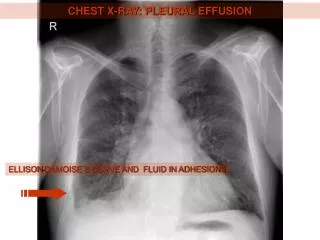
Following CXR shows complete opacity of the lower 2/3 of right chest • The opacity forms a sharp edge shadow with the RUL and extends lateral to the RUL • The sharp edge shadow indicates a pleural surface, placing the opacity outside lung, in pleural space • This is a large pleural effusion, showing what is essentially a high meniscus.
Following CXR is same patient as preceding, but following a thoracentesis • Right effusion is much smaller, but not gone, and there is now pneumothorax as well, a hydropneumthorax (note the air-fluid levels) • In spite of drainage of most of the effusion, there is still nodular thickening of the right pleural surfaces, secondary to tumor implants in this patient with malignant mesothelioma, secondary to prior asbestos exposure
Another CXR on same patient shows progression of mesothelioma encasing entire right lung (over a year later)
Following CXR on 70-year-old female patient who complains of shortness of breath climbing one flight of stairs, worsening over last couple weeks. Had been smoker until 5 years ago when she had small MI
History suggests CHF • Findings of CHF on CXR in general • Cardiomegaly (width of heart greater than 50% of width of lungs at widest point, on standard 6-foot upright PA CXR with good inspiration and not rotated) • This is actually assessment of cardiac silhouette, so remember possibility of pericardial effusion • Don’t apply 50% rule without allowing for any non-standard factors • Pleural effusions • Pulmonary vessel enlargement(especially upper lobe vessels on upright CXR) • Pulmonary edema • Interstitial edema (Kerley lines, peribronchial cuffing, fuzzy vessels) • Alveolar edema (symmetrical air-space infiltrates, diffuse or perihilar/bat wing)
On this patient’s CXR (standard upright PA), cardiac silhouette size is borderline (50%) • She has no visible pleural effusion • No visible pulmonary edema (not surprising since she is only symptomatic with exercise) • However, she does have upper lobe vessel enlargement (compare to following CXR which patient had done 2 months before she became symptomatic)
Following image is magnification of upper lobe vessels when patient was asymptomatic • Note typically thin upper lobe vessels seen on upright CXR