Comprehensive Guide to Chest X-Ray Pathological Correlation and Evaluation
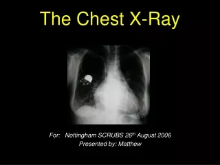
This guide provides an in-depth analysis of chest X-ray (CXR) interpretation focusing on pathological correlations. It covers the identification of normal structures, various densities, and the genesis of abnormal findings. Key concepts include localization of pathologies such as lobar distribution, air bronchograms, silhouette signs, and extrapleural signs. The text also addresses common conditions like consolidation, cavitation, atelectasis, pleural effusion, pneumothorax, and pulmonary edema, detailing evaluation steps for each pathology, with an emphasis on anatomical landmarks and clinical significance.

Comprehensive Guide to Chest X-Ray Pathological Correlation and Evaluation
E N D
Presentation Transcript
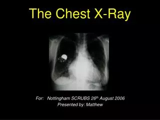
Chest X-ray Path correlation • Normal structures • Densities • Genesis of abnormal densities • Localization • Pathological correlation • Steps in evaluation of CXR
Left RV RA LV LA
Hemorrhage Hem
Localization • Lobar distribution • Air bronchogram • Silouhette sign • Extra pleural sign
LUL RUL RML Lingula RLL LLL Silouhette sign
Peripheral Sharp inner Indistinct outer Concave angles Extrapleural sign
Pathological correlation • Consolidation • Cavitation • Mass • Atelectasis • Pleural effusion • Pneumothorax • Pulmonary edema
Air bronchogram Lobar No loss of volume Consolidation
Hole Wall Lumen Content Cavity
Homogenous Sharp margin No respect Mass
Loss of lung volume Atelectasis
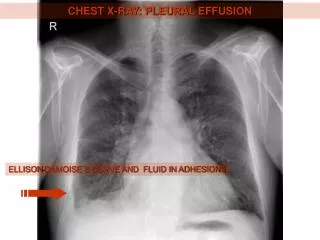
Homogenous Costophrenic angle Meniscus Pleural effusion
Air in pleura Atelectatic lung Hemithorax Mediastinual shift Pneumothorax
Bilateral Diffuse Alveolar Pulmonary edema
Bleb Emphysema