Olfactory Nerve
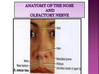
Olfactory Nerve. Dr. Nimir Dr. Safaa. Objectives Describe the formation of the olfactory nerve. Discuss the olfactory pathway. Olfactory Nerves (Cranial Nerve I): It arise from the olfactory receptor nerve cells in olfactory mucous membrane in upper part of nasal cavity.


Olfactory Nerve
E N D
Presentation Transcript
Olfactory Nerve Dr. Nimir Dr. Safaa
Objectives • Describe the formation of the olfactory nerve. • Discuss the olfactory pathway.
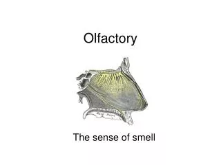
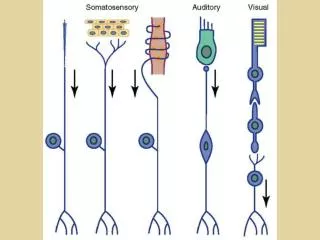
Olfactory Nerves (Cranial Nerve I): • It arise from the olfactory receptor nerve cells in olfactory mucous membrane in upper part of nasal cavity. • Olfactory receptor cells scattered among supporting cells. • Each receptor cell consists of a small bipolar nerve cell with a coarse peripheral process that passes to mucous membrane and a fine central process.
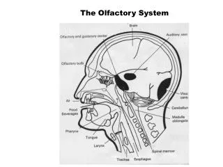
From peripheral process short cilia arise(olfactory hairs) which project into mucus covering mucous membrane. These cilia react to odors in air and stimulate the olfactory cells. • Fine central processes form olfactory nerve fibers. Bundles of these fibers pass through cribriform plate of ethmoid bone to enter the olfactory bulb. The olfactory nerve fibers are unmyelinated and are covered with Schwann cells.
Olfactory Bulb: • It is ovoid structure with several types of nerve cells largest of which is the mitral cell. • Incoming olfactory fibers synapse with dendrites of mitral cells and form rounded areas known as synaptic glomeruli. • Smaller nerve cells, called tufted cells and granular cells, also synapse with mitral cells. • Olfactory bulb, in addition, receives axons from contralateral olfactory bulb through olfactory tract.
Olfactory Tract: • It is narrow band of white matter runs from posterior end of olfactory bulb beneath inferior surface of frontal lobe. • It consists of central axons of mitral and tufted cells of bulb and some centrifugal fibers from the opposite olfactory bulb.
At anterior perforated substance the tract divides into medial and lateral olfactory striae. • Lateral stria carries axons to olfactory area of cerebral cortex (periamygdaloid and prepiriform areas). • Medial olfactory stria carries fibers that cross median plane in anterior commissure to pass to olfactory bulb of opposite side.
Periamygdaloid and prepiriform areas are known as primary olfactory cortex. • Entorhinal area (area 28) of parahippocampalgyruswhich receives connections from primary olfactory cortex is secondary olfactory cortex. These areas are responsible for appreciation of olfactory sensations.
In contrast to all other sensory pathways olfactory pathway has only two neurons and reaches cerebral cortex without synapsing in thalamus. • Primary olfactory cortex sends nerve fibers to many other centers within brain to establish connections for emotional and autonomic responses to olfactory sensations.